 Movie
Movie Controller

[English] 日本語
 Yorodumi
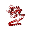
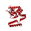
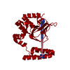
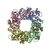
- PDB-1q5o: HCN2J 443-645 in the presence of cAMP, selenomethionine derivative -
+ Open data

- Basic information
| Entry | Database: PDB / ID: 1q5o | ||||||
|---|---|---|---|---|---|---|---|
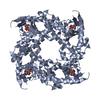
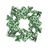
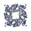
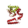
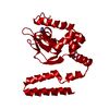
| Title | HCN2J 443-645 in the presence of cAMP, selenomethionine derivative | ||||||
 Components Components | Potassium/sodium hyperpolarization-activated cyclic nucleotide-gated channel 2 | ||||||
 Keywords Keywords | TRANSPORT PROTEIN / CNBD / C-LINKER / PACEMAKER / HCN / HCN2 / CHANNEL / CYCLIC NUCLEOTIDE / CAP / PKA / cAMP / ION CHANNEL / LIGAND | ||||||
| Function / homology |  Function and homology information Function and homology informationHCN channels / cellular response to aldosterone / HCN channel complex / ammonium transmembrane transport / cellular response to cGMP / intracellularly cAMP-activated cation channel activity / sodium ion import across plasma membrane / voltage-gated sodium channel activity / regulation of membrane depolarization / potassium ion import across plasma membrane ...HCN channels / cellular response to aldosterone / HCN channel complex / ammonium transmembrane transport / cellular response to cGMP / intracellularly cAMP-activated cation channel activity / sodium ion import across plasma membrane / voltage-gated sodium channel activity / regulation of membrane depolarization / potassium ion import across plasma membrane / voltage-gated potassium channel activity / cAMP binding / somatodendritic compartment / potassium ion transmembrane transport / dendrite membrane / cellular response to cAMP / dendritic shaft / sodium ion transmembrane transport / PDZ domain binding / regulation of membrane potential / molecular adaptor activity / response to xenobiotic stimulus / axon / neuronal cell body / dendrite / protein-containing complex binding / membrane / identical protein binding / plasma membrane Similarity search - Function | ||||||
| Biological species |  | ||||||
| Method |  X-RAY DIFFRACTION / SYNCHROTRON / MOLECULAR REPLACEMENT / Resolution: 2.3 Å X-RAY DIFFRACTION / SYNCHROTRON / MOLECULAR REPLACEMENT / Resolution: 2.3 Å | ||||||
 Authors Authors | Zagotta, W.N. / Olivier, N.B. / Black, K.D. / Young, E.C. / Olson, R. / Gouaux, J.E. | ||||||
 Citation Citation | Journal: Nature / Year: 2003 Title: STRUCTURAL BASIS FOR MODULATION AND AGONIST SPECIFICITY OF HCN PACEMAKER CHANNELS Authors: Zagotta, W.N. / Olivier, N.B. / Black, K.D. / Young, E.C. / Olson, R. / Gouaux, J.E. | ||||||
| History |
|
- Structure visualization
| Structure viewer | Molecule: MolmilJmol/JSmol |
|---|
- Downloads & links
-Download
| PDBx/mmCIF format | 1q5o.cif.gz | 57.9 KB | Display | PDBx/mmCIF format |
|---|---|---|---|---|
| PDB format | pdb1q5o.ent.gz | 41.4 KB | Display | PDB format |
| PDBx/mmJSON format | 1q5o.json.gz | Tree view | PDBx/mmJSON format | |
| Others |  Other downloads Other downloads |
-Validation report
| Arichive directory | https://data.pdbj.org/pub/pdb/validation_reports/q5/1q5oftp://data.pdbj.org/pub/pdb/validation_reports/q5/1q5o | HTTPS FTP |
|---|
-Related structure data
| Related structure data |  1q3eC  1q43SC C: citing same article ( S: Starting model for refinement |
|---|---|
| Similar structure data |

-Links
 PDBj
PDBj


- Assembly
| Deposited unit | 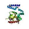
| ||||||||
|---|---|---|---|---|---|---|---|---|---|
| 1 | 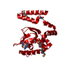
| ||||||||
| Unit cell |
| ||||||||
| Details | The biological assembly is a tetramer. This structure may be generated by applying the symmetry operations: y-1/2,1/2-x,z and -x,1-y,z and 1/2-y,1/2+x,z |
-Components
| #1: Protein | Mass: 24567.943 Da / Num. of mol.: 1 / Fragment: Residues 443-645 Source method: isolated from a genetically manipulated source Source: (gene. exp.)  |
|---|---|
| #2: Chemical | ChemComp-CMP /   Mass: 329.206 Da / Num. of mol.: 1 / Source method: obtained synthetically / Formula: C10H12N5O6P Mass: 329.206 Da / Num. of mol.: 1 / Source method: obtained synthetically / Formula: C10H12N5O6P |
| #3: Water | ChemComp-HOH /  Mass: 18.015 Da / Num. of mol.: 129 / Source method: isolated from a natural source / Formula: H2O Mass: 18.015 Da / Num. of mol.: 129 / Source method: isolated from a natural source / Formula: H2O |
| Has protein modification | Y |
-Experimental details
-Experiment
| Experiment | Method: X-RAY DIFFRACTION / Number of used crystals: 1 |
|---|
- Sample preparation
| Crystal | Density Matthews: 2.32 Å3/Da / Density % sol: 46.96 % | |||||||||||||||||||||||||||||||||||
|---|---|---|---|---|---|---|---|---|---|---|---|---|---|---|---|---|---|---|---|---|---|---|---|---|---|---|---|---|---|---|---|---|---|---|---|---|
| Crystal grow | Temperature: 277 K / Method: vapor diffusion, hanging drop / pH: 4.6 Details: PEG 400, sodium citrate, sodium chloride, DTT, HEPES, cAMP, pH 4.6, VAPOR DIFFUSION, HANGING DROP, temperature 277K | |||||||||||||||||||||||||||||||||||
| Crystal grow | *PLUS Temperature: 4 ℃ / Method: vapor diffusion, hanging drop | |||||||||||||||||||||||||||||||||||
| Components of the solutions | *PLUS
|
-Data collection
| Diffraction | Mean temperature: 100 K |
|---|---|
| Diffraction source | Source: SYNCHROTRON / Site: NSLS  / Beamline: X4A / Wavelength: 0.96429 Å / Beamline: X4A / Wavelength: 0.96429 Å |
| Detector | Type: ADSC QUANTUM 4 / Detector: CCD / Date: Jun 2, 2002 |
| Radiation | Monochromator: SAGITALLY FOCUSED Si(111) / Protocol: MAD / Monochromatic (M) / Laue (L): M / Scattering type: x-ray |
| Radiation wavelength | Wavelength: 0.96429 Å / Relative weight: 1 |
| Reflection | Resolution: 2.1→20 Å / Num. all: 13749 / Num. obs: 13749 / % possible obs: 98 % / Observed criterion σ(F): 0 / Observed criterion σ(I): -3 / Redundancy: 8.5 % / Biso Wilson estimate: 23.85 Å2 / Rmerge(I) obs: 0.069 / Net I/σ(I): 23.62 |
| Reflection shell | Resolution: 2.3→2.44 Å / % possible all: 99.5 |
| Reflection | *PLUS |
- Processing
| Software |
| ||||||||||||||||||||||||||||||||||||
|---|---|---|---|---|---|---|---|---|---|---|---|---|---|---|---|---|---|---|---|---|---|---|---|---|---|---|---|---|---|---|---|---|---|---|---|---|---|
| Refinement | Method to determine structure: MOLECULAR REPLACEMENT Starting model: PDB ENTRY 1Q43 Resolution: 2.3→20 Å / Cross valid method: THROUGHOUT / σ(F): 0 / Stereochemistry target values: Engh & Huber
| ||||||||||||||||||||||||||||||||||||
| Displacement parameters |
| ||||||||||||||||||||||||||||||||||||
| Refine analyze |
| ||||||||||||||||||||||||||||||||||||
| Refinement step | Cycle: LAST / Resolution: 2.3→20 Å
| ||||||||||||||||||||||||||||||||||||
| Refine LS restraints |
| ||||||||||||||||||||||||||||||||||||
| LS refinement shell | Resolution: 2.3→2.44 Å / Rfactor Rfree error: 0.033
| ||||||||||||||||||||||||||||||||||||
| Xplor file |
| ||||||||||||||||||||||||||||||||||||
| Refinement | *PLUS | ||||||||||||||||||||||||||||||||||||
| Solvent computation | *PLUS | ||||||||||||||||||||||||||||||||||||
| Displacement parameters | *PLUS | ||||||||||||||||||||||||||||||||||||
| Refine LS restraints | *PLUS
|
