[English] 日本語
Yorodumi
- PDB-6z6d: Crystal structure of the HK97 bacteriophage large terminase -

+
Open data


ID or keywords:

Loading...

-
Basic information

Entry
Database: PDB / ID: 6z6d
TitleCrystal structure of the HK97 bacteriophage large terminase
ComponentsTerminase large subunit
KeywordsVIRAL PROTEIN / genome packaging / bacteriophage / ATPase / nuclease
Function / homologyTerminase large subunit-like / Terminase large subunit, ATPase domain / BROMIDE ION / Terminase large subunit
Function and homology information
Biological speciesEnterobacteria phage HK97 (virus)
MethodX-RAY DIFFRACTION / SYNCHROTRON / MAD / Resolution: 2.2 Å
AuthorsFung, H.K.H. / Chechik, M. / Baumann, C.G. / Antson, A.A.
Funding support United Kingdom, 2items
OrganizationGrant numberCountry
Wellcome Trust098230 United Kingdom
Wellcome Trust095024MA United Kingdom
Citation
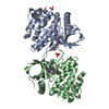
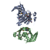
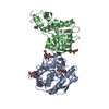
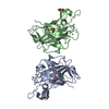
Journal: Nucleic Acids Res / Year: 2022
Title: Structural basis of DNA packaging by a ring-type ATPase from an archetypal viral system.
Authors: Herman K H Fung / Shelley Grimes / Alexis Huet / Robert L Duda / Maria Chechik / Joseph Gault / Carol V Robinson / Roger W Hendrix / Paul J Jardine / James F Conway / Christoph G Baumann / Alfred A Antson /
Abstract: Many essential cellular processes rely on substrate rotation or translocation by a multi-subunit, ring-type NTPase. A large number of double-stranded DNA viruses, including tailed bacteriophages and ...Many essential cellular processes rely on substrate rotation or translocation by a multi-subunit, ring-type NTPase. A large number of double-stranded DNA viruses, including tailed bacteriophages and herpes viruses, use a homomeric ring ATPase to processively translocate viral genomic DNA into procapsids during assembly. Our current understanding of viral DNA packaging comes from three archetypal bacteriophage systems: cos, pac and phi29. Detailed mechanistic understanding exists for pac and phi29, but not for cos. Here, we reconstituted in vitro a cos packaging system based on bacteriophage HK97 and provided a detailed biochemical and structural description. We used a photobleaching-based, single-molecule assay to determine the stoichiometry of the DNA-translocating ATPase large terminase. Crystal structures of the large terminase and DNA-recruiting small terminase, a first for a biochemically defined cos system, reveal mechanistic similarities between cos and pac systems. At the same time, mutational and biochemical analyses indicate a new regulatory mechanism for ATPase multimerization and coordination in the HK97 system. This work therefore establishes a framework for studying the evolutionary relationships between ATP-dependent DNA translocation machineries in double-stranded DNA viruses.
History
DepositionMay 28, 2020Deposition site: PDBE / Processing site: PDBE
Revision 1.0Jun 9, 2021Provider: repository / Type: Initial release
Revision 1.1Dec 21, 2022Group: Database references / Category: citation / citation_author / database_2
Item: _citation.country / _citation.journal_abbrev ..._citation.country / _citation.journal_abbrev / _citation.journal_id_ASTM / _citation.journal_id_CSD / _citation.journal_id_ISSN / _citation.journal_volume / _citation.page_first / _citation.page_last / _citation.pdbx_database_id_DOI / _citation.pdbx_database_id_PubMed / _citation.title / _citation.year / _citation_author.identifier_ORCID / _database_2.pdbx_DOI / _database_2.pdbx_database_accession
Revision 1.2Jun 19, 2024Group: Data collection / Category: chem_comp_atom / chem_comp_bond

-
Structure visualization

Structure viewerMolecule:
MolmilJmol/JSmol

Downloads & links

-
Assembly

Deposited unit
A: Terminase large subunit
hetero molecules


Theoretical massNumber of molelcules
Total (without water)57,80918
Polymers56,4501
Non-polymers1,35817
Water1,67593
1


  • Idetical with deposited unit
  • defined by author&software
  • Evidence: light scattering
TypeNameSymmetry operationNumber
identity operation1_555x,y,z1
Buried area2310 Å2
ΔGint-3 kcal/mol
Surface area20910 Å2
MethodPISA
Unit cell
Length a, b, c (Å)211.787, 39.146, 65.450
Angle α, β, γ (deg.)90.000, 103.390, 90.000
Int Tables number5
Space group name H-MC121

-
Components

#1: Protein Terminase large subunit


Mass: 56450.344 Da / Num. of mol.: 1
Source method: isolated from a genetically manipulated source
Source: (gene. exp.) Enterobacteria phage HK97 (virus) / Production host: Escherichia coli (E. coli) / References: UniProt: Q9MCT1
#2: Chemical
ChemComp-BR / BROMIDE ION


Mass: 79.904 Da / Num. of mol.: 17 / Source method: obtained synthetically / Formula: Br
#3: Water ChemComp-HOH / water


Mass: 18.015 Da / Num. of mol.: 93 / Source method: isolated from a natural source / Formula: H2O
Has ligand of interestN

-
Experimental details

-
Experiment

ExperimentMethod: X-RAY DIFFRACTION / Number of used crystals: 1

-
Sample preparation

CrystalDensity Matthews: 2.34 Å3/Da / Density % sol: 47.39 %
Crystal growTemperature: 293 K / Method: vapor diffusion, hanging drop / Details: 1.6 M ammonium sulfate, pH 7.6

-
Data collection

DiffractionMean temperature: 100 K / Serial crystal experiment: N
Diffraction sourceSource: SYNCHROTRON / Site: Diamond / Beamline: I02 / Wavelength: 0.9198 Å
DetectorType: DECTRIS PILATUS 6M / Detector: PIXEL / Date: Mar 6, 2016
RadiationProtocol: SINGLE WAVELENGTH / Monochromatic (M) / Laue (L): M / Scattering type: x-ray
Radiation wavelengthWavelength: 0.9198 Å / Relative weight: 1
ReflectionResolution: 2.2→103.23 Å / Num. obs: 26796 / % possible obs: 99.6 % / Redundancy: 3.3 % / CC1/2: 0.991 / Rmerge(I) obs: 0.136 / Net I/σ(I): 7.1
Reflection shellResolution: 2.2→2.27 Å / Rmerge(I) obs: 0.842 / Mean I/σ(I) obs: 1.8 / Num. unique obs: 2242 / CC1/2: 0.595 / % possible all: 98.2

-
Processing

Software
NameVersionClassification
REFMAC5.8.0158refinement
PDB_EXTRACT3.22data extraction
XDSOct 15, 2015data reduction
XDSOct 15, 2015data scaling
SHELXD2013/2phasing
RefinementMethod to determine structure: MAD / Resolution: 2.2→103.23 Å / Cor.coef. Fo:Fc: 0.95 / Cor.coef. Fo:Fc free: 0.913 / SU B: 8.595 / SU ML: 0.199 / Cross valid method: THROUGHOUT / σ(F): 0 / ESU R: 0.264 / ESU R Free: 0.215 / Stereochemistry target values: MAXIMUM LIKELIHOOD
Details: HYDROGENS HAVE BEEN ADDED IN THE RIDING POSITIONS U VALUES : REFINED INDIVIDUALLY
RfactorNum. reflection% reflectionSelection details
Rfree0.2542 1244 4.6 %RANDOM
Rwork0.2051 ---
obs0.2072 25551 99.55 %-
Solvent computationIon probe radii: 0.8 Å / Shrinkage radii: 0.8 Å / VDW probe radii: 1.2 Å / Solvent model: MASK
Displacement parametersBiso max: 106.49 Å2 / Biso mean: 36.887 Å2 / Biso min: 16.64 Å2
Baniso -1Baniso -2Baniso -3
1--1.69 Å20 Å2-1.69 Å2
2---1.01 Å20 Å2
3---3.14 Å2
Refinement stepCycle: final / Resolution: 2.2→103.23 Å
ProteinNucleic acidLigandSolventTotal
Num. atoms3522 0 18 93 3633
Biso mean--66.24 33.34 -
Num. residues----479
Refine LS restraints
Refine-IDTypeDev idealDev ideal targetNumber
X-RAY DIFFRACTIONr_bond_refined_d0.0080.0193591
X-RAY DIFFRACTIONr_bond_other_d0.0010.023316
X-RAY DIFFRACTIONr_angle_refined_deg1.2581.964894
X-RAY DIFFRACTIONr_angle_other_deg0.90537647
X-RAY DIFFRACTIONr_dihedral_angle_1_deg7.8015.042479
X-RAY DIFFRACTIONr_dihedral_angle_2_deg38.6524.526137
X-RAY DIFFRACTIONr_dihedral_angle_3_deg12.08615549
X-RAY DIFFRACTIONr_dihedral_angle_4_deg12.9131518
X-RAY DIFFRACTIONr_chiral_restr0.0640.2576
X-RAY DIFFRACTIONr_gen_planes_refined0.0070.0214064
X-RAY DIFFRACTIONr_gen_planes_other0.0010.02698
LS refinement shellResolution: 2.204→2.261 Å / Rfactor Rfree error: 0 / Total num. of bins used: 20
RfactorNum. reflection% reflection
Rfree0.335 88 -
Rwork0.33 1820 -
all-1908 -
obs--97.85 %

+
About Yorodumi

-
News

-
Feb 9, 2022. New format data for meta-information of EMDB entries

New format data for meta-information of EMDB entries

  • Version 3 of the EMDB header file is now the official format.
  • The previous official version 1.9 will be removed from the archive.

Related info.:EMDB header

External links:wwPDB to switch to version 3 of the EMDB data model

-
Aug 12, 2020. Covid-19 info

Covid-19 info

URL: https://pdbjlvh1.pdbj.org/emnavi/covid19.php

New page: Covid-19 featured information page in EM Navigator.

Related info.:Covid-19 info / Mar 5, 2020. Novel coronavirus structure data

+
Mar 5, 2020. Novel coronavirus structure data

Novel coronavirus structure data

Related info.:Yorodumi Speices / Aug 12, 2020. Covid-19 info

External links:COVID-19 featured content - PDBj / Molecule of the Month (242):Coronavirus Proteases

+
Jan 31, 2019. EMDB accession codes are about to change! (news from PDBe EMDB page)

EMDB accession codes are about to change! (news from PDBe EMDB page)

  • The allocation of 4 digits for EMDB accession codes will soon come to an end. Whilst these codes will remain in use, new EMDB accession codes will include an additional digit and will expand incrementally as the available range of codes is exhausted. The current 4-digit format prefixed with “EMD-” (i.e. EMD-XXXX) will advance to a 5-digit format (i.e. EMD-XXXXX), and so on. It is currently estimated that the 4-digit codes will be depleted around Spring 2019, at which point the 5-digit format will come into force.
  • The EM Navigator/Yorodumi systems omit the EMD- prefix.

Related info.:Q: What is EMD? / ID/Accession-code notation in Yorodumi/EM Navigator

External links:EMDB Accession Codes are Changing Soon! / Contact to PDBj

+
Jul 12, 2017. Major update of PDB

Major update of PDB

  • wwPDB released updated PDB data conforming to the new PDBx/mmCIF dictionary.
  • This is a major update changing the version number from 4 to 5, and with Remediation, in which all the entries are updated.
  • In this update, many items about electron microscopy experimental information are reorganized (e.g. em_software).
  • Now, EM Navigator and Yorodumi are based on the updated data.

External links:wwPDB Remediation / Enriched Model Files Conforming to OneDep Data Standards Now Available in the PDB FTP Archive

-
Yorodumi

Thousand views of thousand structures

  • Yorodumi is a browser for structure data from EMDB, PDB, SASBDB, etc.
  • This page is also the successor to EM Navigator detail page, and also detail information page/front-end page for Omokage search.
  • The word "yorodu" (or yorozu) is an old Japanese word meaning "ten thousand". "mi" (miru) is to see.

Related info.:EMDB / PDB / SASBDB / Comparison of 3 databanks / Yorodumi Search / Aug 31, 2016. New EM Navigator & Yorodumi / Yorodumi Papers / Jmol/JSmol / Function and homology information / Changes in new EM Navigator and Yorodumi

Read more