[English] 日本語
Yorodumi
- PDB-4d7u: The structure of the catalytic domain of NcLPMO9C from the filame... -

+
Open data


ID or keywords:

Loading...

-
Basic information

Entry
Database: PDB / ID: 4d7u
Title
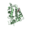
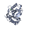
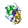
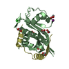
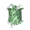
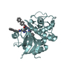
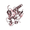
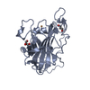
The structure of the catalytic domain of NcLPMO9C from the filamentous fungus Neurospora crassa
ComponentsENDOGLUCANASE II
KeywordsOXIDOREDUCTASE / LYTIC MONOOXYGENASE / HEMICELLULOSE ACTIVE / AA9
Function / homology
Function and homology information


cellulose binding / carbohydrate metabolic process / extracellular region / metal ion binding
Similarity search - Function
Coagulation Factor XIII; Chain A, domain 1 - #70 / Auxiliary Activity family 9 / Auxiliary Activity family 9 (formerly GH61) / CBM1 (carbohydrate binding type-1) domain signature. / Cellulose-binding domain, fungal / Cellulose-binding domain superfamily / Fungal cellulose binding domain / CBM1 (carbohydrate binding type-1) domain profile. / Fungal-type cellulose-binding domain / Coagulation Factor XIII; Chain A, domain 1 ...Coagulation Factor XIII; Chain A, domain 1 - #70 / Auxiliary Activity family 9 / Auxiliary Activity family 9 (formerly GH61) / CBM1 (carbohydrate binding type-1) domain signature. / Cellulose-binding domain, fungal / Cellulose-binding domain superfamily / Fungal cellulose binding domain / CBM1 (carbohydrate binding type-1) domain profile. / Fungal-type cellulose-binding domain / Coagulation Factor XIII; Chain A, domain 1 / Distorted Sandwich / Mainly Beta
Similarity search - Domain/homology
COPPER (II) ION / Endoglucanase II
Similarity search - Component
Biological speciesNEUROSPORA CRASSA (fungus)
MethodX-RAY DIFFRACTION / SYNCHROTRON / MOLECULAR REPLACEMENT / Resolution: 1.56 Å
AuthorsBorisova, A.S. / Isaksen, T. / Mathiesen, G. / Sorlie, M. / Sandgren, M. / Eijsink, V.G.H. / Dimarogona, M.
CitationJournal: J.Biol.Chem. / Year: 2015
Title: Structural and Functional Characterization of a Lytic Polysaccharide Monooxygenase with Broad Substrate Specificity
Authors: Borisova, A.S. / Isaksen, T. / Dimarogona, M. / Kognole, A.A. / Mathiesen, G. / Varnai, A. / Rohr, A.K. / Payne, C. / Sorlie, M. / Sandgren, M. / Eijsink, V.G.H. / Sorlie, M.
History
DepositionNov 27, 2014Deposition site: PDBE / Processing site: PDBE
Revision 1.0Jul 22, 2015Provider: repository / Type: Initial release
Revision 1.1Jul 29, 2015Group: Database references
Revision 1.2Oct 7, 2015Group: Database references
Revision 1.3Jan 17, 2018Group: Data collection / Category: diffrn_source / Item: _diffrn_source.pdbx_synchrotron_site
Revision 1.4Dec 20, 2023Group: Data collection / Database references ...Data collection / Database references / Derived calculations / Other / Refinement description
Category: chem_comp_atom / chem_comp_bond ...chem_comp_atom / chem_comp_bond / database_2 / pdbx_database_status / pdbx_initial_refinement_model / struct_conn / struct_site
Item: _database_2.pdbx_DOI / _database_2.pdbx_database_accession ..._database_2.pdbx_DOI / _database_2.pdbx_database_accession / _pdbx_database_status.status_code_sf / _struct_conn.ptnr1_auth_comp_id / _struct_conn.ptnr1_auth_seq_id / _struct_conn.ptnr1_label_asym_id / _struct_conn.ptnr1_label_atom_id / _struct_conn.ptnr1_label_comp_id / _struct_conn.ptnr1_label_seq_id / _struct_conn.ptnr2_auth_comp_id / _struct_conn.ptnr2_auth_seq_id / _struct_conn.ptnr2_label_asym_id / _struct_conn.ptnr2_label_atom_id / _struct_conn.ptnr2_label_comp_id / _struct_conn.ptnr2_label_seq_id / _struct_site.pdbx_auth_asym_id / _struct_site.pdbx_auth_comp_id / _struct_site.pdbx_auth_seq_id

-
Structure visualization

Structure viewerMolecule:
MolmilJmol/JSmol

Downloads & links

-
Assembly

Deposited unit
A: ENDOGLUCANASE II
B: ENDOGLUCANASE II
hetero molecules


Theoretical massNumber of molelcules
Total (without water)47,2789
Polymers46,6902
Non-polymers5887
Water10,052558
1
A: ENDOGLUCANASE II
hetero molecules


Theoretical massNumber of molelcules
Total (without water)23,5934
Polymers23,3451
Non-polymers2483
Water181
TypeNameSymmetry operationNumber
identity operation1_555x,y,z1
MethodPISA
2
B: ENDOGLUCANASE II
hetero molecules


Theoretical massNumber of molelcules
Total (without water)23,6855
Polymers23,3451
Non-polymers3404
Water181
TypeNameSymmetry operationNumber
identity operation1_555x,y,z1
MethodPISA
Unit cell
Length a, b, c (Å)44.258, 66.755, 66.077
Angle α, β, γ (deg.)90.00, 101.26, 90.00
Int Tables number4
Space group name H-MP1211

-
Components

#1: Protein ENDOGLUCANASE II / NCLPMO9C-N


Mass: 23345.139 Da / Num. of mol.: 2 / Fragment: CATALYTIC DOMAIN, RESIDUES 17-243
Source method: isolated from a genetically manipulated source
Source: (gene. exp.) NEUROSPORA CRASSA (fungus) / Strain: OR74A / Plasmid: PPINK-GAPHC / Production host: KOMAGATAELLA PASTORIS (fungus) / Strain (production host): PICHIAPINK STRAIN4 / References: UniProt: Q7SHI8
#2: Chemical ChemComp-CU / COPPER (II) ION


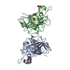
Mass: 63.546 Da / Num. of mol.: 2 / Source method: obtained synthetically / Formula: Cu
#3: Chemical
ChemComp-GOL / GLYCEROL / GLYCERIN / PROPANE-1,2,3-TRIOL


Mass: 92.094 Da / Num. of mol.: 5 / Source method: obtained synthetically / Formula: C3H8O3
#4: Water ChemComp-HOH / water


Mass: 18.015 Da / Num. of mol.: 558 / Source method: isolated from a natural source / Formula: H2O

-
Experimental details

-
Experiment

ExperimentMethod: X-RAY DIFFRACTION / Number of used crystals: 1

-
Sample preparation

CrystalDensity Matthews: 1.99 Å3/Da / Density % sol: 38 % / Description: NONE
Crystal growpH: 5.1 / Details: 0.2M AMMONIUM CITRATE, 20% PEG 3350, PH 5.1

-
Data collection

DiffractionMean temperature: 100 K
Diffraction sourceSource: SYNCHROTRON / Site: MAX II / Beamline: I911-3 / Wavelength: 1
DetectorType: MARMOSAIC 225 mm CCD / Detector: CCD / Date: May 15, 2014 / Details: MIRRORS
RadiationMonochromator: SI(111) / Protocol: SINGLE WAVELENGTH / Monochromatic (M) / Laue (L): M / Scattering type: x-ray
Radiation wavelengthWavelength: 1 Å / Relative weight: 1
ReflectionResolution: 1.56→64.81 Å / Num. obs: 49150 / % possible obs: 91.3 % / Observed criterion σ(I): 2 / Redundancy: 3.3 % / Biso Wilson estimate: 12.6 Å2 / Rmerge(I) obs: 0.04 / Net I/σ(I): 15.5
Reflection shellResolution: 1.56→1.59 Å / Redundancy: 1.8 % / Mean I/σ(I) obs: 3.1 / % possible all: 29.2

-
Processing

Software
NameVersionClassification
REFMAC5.8.0073refinement
XDSdata reduction
Aimlessdata scaling
PHASERphasing
RefinementMethod to determine structure: MOLECULAR REPLACEMENT
Starting model: PDB ENTRY 4EIR
Resolution: 1.56→64.805 Å / Cor.coef. Fo:Fc: 0.971 / Cor.coef. Fo:Fc free: 0.96 / SU B: 1.764 / SU ML: 0.061 / Cross valid method: THROUGHOUT / ESU R: 0.092 / ESU R Free: 0.09 / Stereochemistry target values: MAXIMUM LIKELIHOOD / Details: HYDROGENS HAVE BEEN ADDED IN THE RIDING POSITIONS.
RfactorNum. reflection% reflectionSelection details
Rfree0.1876 2423 5.02 %RANDOM
Rwork0.1563 ---
obs0.158 48932 91.057 %-
Solvent computationIon probe radii: 0.8 Å / Shrinkage radii: 0.8 Å / VDW probe radii: 1.2 Å / Solvent model: MASK BULK SOLVENT
Displacement parametersBiso mean: 13.496 Å2
Baniso -1Baniso -2Baniso -3
1--1.209 Å20 Å2-0.206 Å2
2--2.347 Å20 Å2
3----0.978 Å2
Refinement stepCycle: LAST / Resolution: 1.56→64.805 Å
ProteinNucleic acidLigandSolventTotal
Num. atoms3280 0 32 558 3870
Refine LS restraints
Refine-IDTypeDev idealDev ideal targetNumber
X-RAY DIFFRACTIONr_bond_refined_d0.0050.0193511
X-RAY DIFFRACTIONr_bond_other_d0.0010.023238
X-RAY DIFFRACTIONr_angle_refined_deg1.1581.9384794
X-RAY DIFFRACTIONr_angle_other_deg0.7183.0027480
X-RAY DIFFRACTIONr_dihedral_angle_1_deg6.2315482
X-RAY DIFFRACTIONr_dihedral_angle_2_deg34.9825.878131
X-RAY DIFFRACTIONr_dihedral_angle_3_deg10.48915503
X-RAY DIFFRACTIONr_dihedral_angle_4_deg9.067154
X-RAY DIFFRACTIONr_chiral_restr0.0690.2522
X-RAY DIFFRACTIONr_gen_planes_refined0.0040.0214181
X-RAY DIFFRACTIONr_gen_planes_other0.0010.02783
X-RAY DIFFRACTIONr_nbd_refined0.2240.21236
X-RAY DIFFRACTIONr_nbd_other0.1550.271
X-RAY DIFFRACTIONr_nbtor_refined0.1690.21765
X-RAY DIFFRACTIONr_nbtor_other
X-RAY DIFFRACTIONr_xyhbond_nbd_refined0.0930.291
X-RAY DIFFRACTIONr_xyhbond_nbd_other0.0490.22
X-RAY DIFFRACTIONr_metal_ion_refined0.050.24
X-RAY DIFFRACTIONr_metal_ion_other
X-RAY DIFFRACTIONr_symmetry_vdw_refined
X-RAY DIFFRACTIONr_symmetry_vdw_other
X-RAY DIFFRACTIONr_symmetry_hbond_refined
X-RAY DIFFRACTIONr_symmetry_hbond_other
X-RAY DIFFRACTIONr_symmetry_metal_ion_refined
X-RAY DIFFRACTIONr_symmetry_metal_ion_other
X-RAY DIFFRACTIONr_mcbond_it0.6161.2651892
X-RAY DIFFRACTIONr_mcbond_other0.6161.2641891
X-RAY DIFFRACTIONr_mcangle_it1.1081.8952386
X-RAY DIFFRACTIONr_mcangle_other
X-RAY DIFFRACTIONr_scbond_it0.6071.2651619
X-RAY DIFFRACTIONr_scbond_other
X-RAY DIFFRACTIONr_scangle_it1.0061.8762408
X-RAY DIFFRACTIONr_scangle_other
X-RAY DIFFRACTIONr_long_range_B_refined
X-RAY DIFFRACTIONr_long_range_B_other
X-RAY DIFFRACTIONr_rigid_bond_restr
X-RAY DIFFRACTIONr_sphericity_free
X-RAY DIFFRACTIONr_sphericity_bonded
LS refinement shellResolution: 1.56→1.601 Å / Total num. of bins used: 20
RfactorNum. reflection% reflection
Rfree0.28 71 -
Rwork0.248 1462 -
obs--38.566 %

+
About Yorodumi

-
News

-
Feb 9, 2022. New format data for meta-information of EMDB entries

New format data for meta-information of EMDB entries

  • Version 3 of the EMDB header file is now the official format.
  • The previous official version 1.9 will be removed from the archive.

Related info.:EMDB header

External links:wwPDB to switch to version 3 of the EMDB data model

-
Aug 12, 2020. Covid-19 info

Covid-19 info

URL: https://pdbjlvh1.pdbj.org/emnavi/covid19.php

New page: Covid-19 featured information page in EM Navigator.

Related info.:Covid-19 info / Mar 5, 2020. Novel coronavirus structure data

+
Mar 5, 2020. Novel coronavirus structure data

Novel coronavirus structure data

Related info.:Yorodumi Speices / Aug 12, 2020. Covid-19 info

External links:COVID-19 featured content - PDBj / Molecule of the Month (242):Coronavirus Proteases

+
Jan 31, 2019. EMDB accession codes are about to change! (news from PDBe EMDB page)

EMDB accession codes are about to change! (news from PDBe EMDB page)

  • The allocation of 4 digits for EMDB accession codes will soon come to an end. Whilst these codes will remain in use, new EMDB accession codes will include an additional digit and will expand incrementally as the available range of codes is exhausted. The current 4-digit format prefixed with “EMD-” (i.e. EMD-XXXX) will advance to a 5-digit format (i.e. EMD-XXXXX), and so on. It is currently estimated that the 4-digit codes will be depleted around Spring 2019, at which point the 5-digit format will come into force.
  • The EM Navigator/Yorodumi systems omit the EMD- prefix.

Related info.:Q: What is EMD? / ID/Accession-code notation in Yorodumi/EM Navigator

External links:EMDB Accession Codes are Changing Soon! / Contact to PDBj

+
Jul 12, 2017. Major update of PDB

Major update of PDB

  • wwPDB released updated PDB data conforming to the new PDBx/mmCIF dictionary.
  • This is a major update changing the version number from 4 to 5, and with Remediation, in which all the entries are updated.
  • In this update, many items about electron microscopy experimental information are reorganized (e.g. em_software).
  • Now, EM Navigator and Yorodumi are based on the updated data.

External links:wwPDB Remediation / Enriched Model Files Conforming to OneDep Data Standards Now Available in the PDB FTP Archive

-
Yorodumi

Thousand views of thousand structures

  • Yorodumi is a browser for structure data from EMDB, PDB, SASBDB, etc.
  • This page is also the successor to EM Navigator detail page, and also detail information page/front-end page for Omokage search.
  • The word "yorodu" (or yorozu) is an old Japanese word meaning "ten thousand". "mi" (miru) is to see.

Related info.:EMDB / PDB / SASBDB / Comparison of 3 databanks / Yorodumi Search / Aug 31, 2016. New EM Navigator & Yorodumi / Yorodumi Papers / Jmol/JSmol / Function and homology information / Changes in new EM Navigator and Yorodumi

Read more