 Movie
Movie Controller

[English] 日本語
 Yorodumi
- PDB-4d7v: The structure of the catalytic domain of NcLPMO9C from the filame... -
+ Open data

- Basic information
| Entry | Database: PDB / ID: 4d7v | ||||||
|---|---|---|---|---|---|---|---|
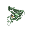
| Title | The structure of the catalytic domain of NcLPMO9C from the filamentous fungus Neurospora crassa | ||||||
 Components Components | ENDOGLUCANASE II | ||||||
 Keywords Keywords | OXIDOREDUCTASE / CATALYTIC DOMAIN / HEMICELLULOSE ACTIVE / AA9 | ||||||
| Function / homology |  Function and homology information Function and homology informationlytic cellulose monooxygenase (C4-dehydrogenating) / cellulose binding / cellulose catabolic process / monooxygenase activity / extracellular region / metal ion binding Similarity search - Function | ||||||
| Biological species |  NEUROSPORA CRASSA (fungus) NEUROSPORA CRASSA (fungus) | ||||||
| Method |  X-RAY DIFFRACTION / SYNCHROTRON / MOLECULAR REPLACEMENT / Resolution: 1.9 Å X-RAY DIFFRACTION / SYNCHROTRON / MOLECULAR REPLACEMENT / Resolution: 1.9 Å | ||||||
 Authors Authors | Borisova, A.S. / Isaksen, T. / Sandgren, M. / Sorlie, M. / Eijsink, V.G.H. / Dimarogona, M. | ||||||
 Citation Citation | Journal: J.Biol.Chem. / Year: 2015 Title: Structural and Functional Characterization of a Lytic Polysaccharide Monooxygenase with Broad Substrate Specificity Authors: Borisova, A.S. / Isaksen, T. / Dimarogona, M. / Kognole, A.A. / Mathiesen, G. / Varnai, A. / Rohr, A.K. / Payne, C. / Sorlie, M. / Sandgren, M. / Eijsink, V.G.H. / Sorlie, M. | ||||||
| History |
|
- Structure visualization
| Structure viewer | Molecule: MolmilJmol/JSmol |
|---|
- Downloads & links
-Download
| PDBx/mmCIF format | 4d7v.cif.gz | 107 KB | Display | PDBx/mmCIF format |
|---|---|---|---|---|
| PDB format | pdb4d7v.ent.gz | 81.9 KB | Display | PDB format |
| PDBx/mmJSON format | 4d7v.json.gz | Tree view | PDBx/mmJSON format | |
| Others |  Other downloads Other downloads |
-Validation report
| Arichive directory | https://data.pdbj.org/pub/pdb/validation_reports/d7/4d7vftp://data.pdbj.org/pub/pdb/validation_reports/d7/4d7v | HTTPS FTP |
|---|
-Related structure data


-Links
 PDBj
PDBj- Assembly
| Deposited unit | 
| ||||||||
|---|---|---|---|---|---|---|---|---|---|
| 1 | 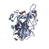
| ||||||||
| 2 | 
| ||||||||
| Unit cell |
|
-Components
| #1: Protein | Mass: 23345.139 Da / Num. of mol.: 2 / Fragment: AA9, RESIDUES 17-243 Source method: isolated from a genetically manipulated source Details: CATALYTIC DOMAIN OF NCLPMO9C Source: (gene. exp.) NEUROSPORA CRASSA (STRAIN ATCC 24698 / 74-OR23-1A / CBS 708.71 / DSM 1257 / FGSC 987) (fungus)Strain: ATCC 24698 / 74-OR23-1A / CBS 708.71 / DSM 1257 / FGSC 987 Gene: GH61-3, NCU02916 / Plasmid: PPINK-GAPHC / Production host: PICHIA PASTORIS (fungus) / Strain (production host): PICHIAPINK STRAIN4 / References: UniProt: Q7SHI8#2: Chemical | ChemComp-ZN /   Mass: 65.409 Da / Num. of mol.: 6 / Source method: obtained synthetically / Formula: Zn Mass: 65.409 Da / Num. of mol.: 6 / Source method: obtained synthetically / Formula: Zn#3: Chemical | ChemComp-ACT /   Mass: 59.044 Da / Num. of mol.: 8 / Source method: obtained synthetically / Formula: C2H3O2 Mass: 59.044 Da / Num. of mol.: 8 / Source method: obtained synthetically / Formula: C2H3O2#4: Chemical |   Mass: 92.094 Da / Num. of mol.: 2 / Source method: obtained synthetically / Formula: C3H8O3 Mass: 92.094 Da / Num. of mol.: 2 / Source method: obtained synthetically / Formula: C3H8O3#5: Water | ChemComp-HOH / |  Mass: 18.015 Da / Num. of mol.: 423 / Source method: isolated from a natural source / Formula: H2O Mass: 18.015 Da / Num. of mol.: 423 / Source method: isolated from a natural source / Formula: H2OHas protein modification | Y | Sequence details | CATALYTIC DOMAIN OF FULL LENGTH ENZYME | |
|---|
-Experimental details
-Experiment
| Experiment | Method: X-RAY DIFFRACTION / Number of used crystals: 1 |
|---|
- Sample preparation
| Crystal | Density Matthews: 1.93 Å3/Da / Density % sol: 36.4 % / Description: NONE |
|---|---|
| Crystal grow | pH: 8 / Details: 0.2M ZINC ACETATE, 17.5% PEG3350, pH 8 |
-Data collection
| Diffraction | Mean temperature: 100 K |
|---|---|
| Diffraction source | Source: SYNCHROTRON / Site: MAX II  / Beamline: I911-3 / Wavelength: 1 / Beamline: I911-3 / Wavelength: 1 |
| Detector | Type: MARMOSAIC 225 mm CCD / Detector: CCD / Date: Dec 15, 2013 / Details: MIRRORS |
| Radiation | Monochromator: SI(111) / Protocol: SINGLE WAVELENGTH / Monochromatic (M) / Laue (L): M / Scattering type: x-ray |
| Radiation wavelength | Wavelength: 1 Å / Relative weight: 1 |
| Reflection | Resolution: 1.9→64.41 Å / Num. obs: 29831 / % possible obs: 99.3 % / Observed criterion σ(I): 2 / Redundancy: 6.7 % / Biso Wilson estimate: 7.5 Å2 / Rmerge(I) obs: 0.31 / Net I/σ(I): 6.8 |
| Reflection shell | Resolution: 1.9→1.94 Å / Redundancy: 6.6 % / Rmerge(I) obs: 1.27 / Mean I/σ(I) obs: 2 / % possible all: 98.4 |
- Processing
| Software |
| ||||||||||||||||||||||||||||||||||||||||||||||||||||||||||||||||||||||||||||||||||||||||||||||||||||||||||||||||||||||||||||||||||||||||||||||||||||||||||||||||||||||||||||||||||||||
|---|---|---|---|---|---|---|---|---|---|---|---|---|---|---|---|---|---|---|---|---|---|---|---|---|---|---|---|---|---|---|---|---|---|---|---|---|---|---|---|---|---|---|---|---|---|---|---|---|---|---|---|---|---|---|---|---|---|---|---|---|---|---|---|---|---|---|---|---|---|---|---|---|---|---|---|---|---|---|---|---|---|---|---|---|---|---|---|---|---|---|---|---|---|---|---|---|---|---|---|---|---|---|---|---|---|---|---|---|---|---|---|---|---|---|---|---|---|---|---|---|---|---|---|---|---|---|---|---|---|---|---|---|---|---|---|---|---|---|---|---|---|---|---|---|---|---|---|---|---|---|---|---|---|---|---|---|---|---|---|---|---|---|---|---|---|---|---|---|---|---|---|---|---|---|---|---|---|---|---|---|---|---|---|
| Refinement | Method to determine structure: MOLECULAR REPLACEMENT Starting model: SAME PROTEIN WITH ONE METAL INSTEAD OF 3 Resolution: 1.9→64.406 Å / Cor.coef. Fo:Fc: 0.942 / Cor.coef. Fo:Fc free: 0.906 / SU B: 5.829 / SU ML: 0.165 / Cross valid method: THROUGHOUT / ESU R: 0.223 / ESU R Free: 0.19 / Stereochemistry target values: MAXIMUM LIKELIHOOD Details: HYDROGENS HAVE BEEN ADDED IN THE RIDING POSITIONS. RESIDUES 182-184 HAVE BEEN OMITTED IN CHAINB DUE TO INSUFFICIENT ELECTRON DENSITY
| ||||||||||||||||||||||||||||||||||||||||||||||||||||||||||||||||||||||||||||||||||||||||||||||||||||||||||||||||||||||||||||||||||||||||||||||||||||||||||||||||||||||||||||||||||||||
| Solvent computation | Ion probe radii: 0.8 Å / Shrinkage radii: 0.8 Å / VDW probe radii: 1.2 Å / Solvent model: MASK BULK SOLVENT | ||||||||||||||||||||||||||||||||||||||||||||||||||||||||||||||||||||||||||||||||||||||||||||||||||||||||||||||||||||||||||||||||||||||||||||||||||||||||||||||||||||||||||||||||||||||
| Displacement parameters | Biso mean: 9.993 Å2
| ||||||||||||||||||||||||||||||||||||||||||||||||||||||||||||||||||||||||||||||||||||||||||||||||||||||||||||||||||||||||||||||||||||||||||||||||||||||||||||||||||||||||||||||||||||||
| Refinement step | Cycle: LAST / Resolution: 1.9→64.406 Å
| ||||||||||||||||||||||||||||||||||||||||||||||||||||||||||||||||||||||||||||||||||||||||||||||||||||||||||||||||||||||||||||||||||||||||||||||||||||||||||||||||||||||||||||||||||||||
| Refine LS restraints |
|