SEQUENCE CLONING ARTIFACT. THE CONSTRUCT WAS PCR AMPLIFIED WITH TAQ POLYMERASE. SEQUENCING OF THE ...SEQUENCE CLONING ARTIFACT. THE CONSTRUCT WAS PCR AMPLIFIED WITH TAQ POLYMERASE. SEQUENCING OF THE CLONED CONSTRUCT INDICATED THAT VAL IN POSITION 161 WAS MUTATED TO ILE.
Resolution: 1.85→50 Å / Num. obs: 42225 / % possible obs: 99.77 % / Redundancy: 3.89 % / Biso Wilson estimate: 27.53 Å2 / Rsym value: 0.069 / Net I/σ(I): 17.88
Reflection shell
Resolution: 1.85→1.92 Å / Redundancy: 3.57 % / Mean I/σ(I) obs: 3.57 / Num. unique all: 4159 / Rsym value: 0.343 / % possible all: 98.39
-
Processing
Software
Name
Version
Classification
DENZO
datareduction
SCALEPACK
datascaling
SOLVE
phasing
RESOLVE
modelbuilding
REFMAC
5.1.9999
refinement
RESOLVE
phasing
Refinement
Method to determine structure: MAD / Resolution: 1.85→37.44 Å / Cor.coef. Fo:Fc: 0.968 / Cor.coef. Fo:Fc free: 0.947 / SU B: 5.233 / SU ML: 0.082 / TLS residual ADP flag: LIKELY RESIDUAL / Cross valid method: THROUGHOUT / ESU R: 0.127 / ESU R Free: 0.123 / Stereochemistry target values: MAXIMUM LIKELIHOOD Details: 1. HYDROGENS HAVE BEEN ADDED IN THE RIDING POSITIONS 2. NON-ROTOMERS: LEU 156 AND LEU 160 (MOLECULE A AND B).
Rfactor
Num. reflection
% reflection
Selection details
Rfree
0.1894
2060
5 %
RANDOM
Rwork
0.14292
-
-
-
obs
0.14523
39479
100 %
-
Solvent computation
Ion probe radii: 0.8 Å / Shrinkage radii: 0.8 Å / VDW probe radii: 1.2 Å / Solvent model: BABINET MODEL WITH MASK
Displacement parameters
Biso mean: 26.642 Å2
Baniso -1
Baniso -2
Baniso -3
1-
-0.07 Å2
0 Å2
-0.02 Å2
2-
-
0.07 Å2
0 Å2
3-
-
-
-0.01 Å2
Refinement step
Cycle: LAST / Resolution: 1.85→37.44 Å
Protein
Nucleic acid
Ligand
Solvent
Total
Num. atoms
4133
0
13
454
4600
Refine LS restraints
Refine-ID
Type
Dev ideal
Dev ideal target
Number
X-RAY DIFFRACTION
r_bond_refined_d
0.02
0.022
4282
X-RAY DIFFRACTION
r_bond_other_d
0.003
0.02
3868
X-RAY DIFFRACTION
r_angle_refined_deg
1.698
1.955
5801
X-RAY DIFFRACTION
r_angle_other_deg
0.909
3
9055
X-RAY DIFFRACTION
r_dihedral_angle_1_deg
5.771
5
505
X-RAY DIFFRACTION
r_dihedral_angle_2_deg
40.422
25.271
203
X-RAY DIFFRACTION
r_dihedral_angle_3_deg
13.328
15
782
X-RAY DIFFRACTION
r_dihedral_angle_4_deg
16.53
15
16
X-RAY DIFFRACTION
r_chiral_restr
0.11
0.2
669
X-RAY DIFFRACTION
r_gen_planes_refined
0.008
0.02
4625
X-RAY DIFFRACTION
r_gen_planes_other
0.001
0.02
811
X-RAY DIFFRACTION
r_nbd_refined
0.215
0.2
955
X-RAY DIFFRACTION
r_nbd_other
0.181
0.2
4037
X-RAY DIFFRACTION
r_nbtor_other
0.088
0.2
2356
X-RAY DIFFRACTION
r_xyhbond_nbd_refined
0.17
0.2
424
X-RAY DIFFRACTION
r_symmetry_vdw_refined
0.245
0.2
26
X-RAY DIFFRACTION
r_symmetry_vdw_other
0.266
0.2
71
X-RAY DIFFRACTION
r_symmetry_hbond_refined
0.237
0.2
31
X-RAY DIFFRACTION
r_mcbond_it
1.496
1.5
2801
X-RAY DIFFRACTION
r_mcbond_other
0.373
1.5
1030
X-RAY DIFFRACTION
r_mcangle_it
1.659
2
4214
X-RAY DIFFRACTION
r_scbond_it
3.072
3
1876
X-RAY DIFFRACTION
r_scangle_it
4.247
4.5
1587
LS refinement shell
Resolution: 1.852→1.9 Å / Total num. of bins used: 20
Rfactor
Num. reflection
% reflection
Rfree
0.264
127
4.92 %
Rwork
0.174
2453
-
Refinement TLS params.
Method: refined / Refine-ID: X-RAY DIFFRACTION
ID
L11 (°2)
L12 (°2)
L13 (°2)
L22 (°2)
L23 (°2)
L33 (°2)
S11 (Å °)
S12 (Å °)
S13 (Å °)
S21 (Å °)
S22 (Å °)
S23 (Å °)
S31 (Å °)
S32 (Å °)
S33 (Å °)
T11 (Å2)
T12 (Å2)
T13 (Å2)
T22 (Å2)
T23 (Å2)
T33 (Å2)
Origin x (Å)
Origin y (Å)
Origin z (Å)
1
0.672
0.1523
-0.0172
0.5367
-0.0587
1.179
-0.0142
-0.0893
-0.0363
0.0354
-0.0006
-0.0654
0.0542
-0.0167
0.0147
-0.1074
-0.001
0.0052
-0.2667
-0.0023
-0.2105
-8.685
31.714
73.862
2
0.9365
0.0487
0.0372
0.8011
0.1341
1.7341
0.0503
0.131
-0.0146
-0.0491
0.0193
-0.0483
-0.0038
-0.0144
-0.0696
-0.1262
-0.0111
0.013
-0.2414
0.0089
-0.2004
5.075
37.61
41.835
Refinement TLS group
Refine-ID: X-RAY DIFFRACTION / Selection: ALL
ID
Refine TLS-ID
Auth asym-ID
Label asym-ID
Auth seq-ID
Label seq-ID
1
1
A
A
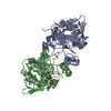
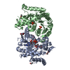
7 - 261
19 - 273
2
2
B
B
10 - 261
22 - 273
+
About Yorodumi
-
News
-
Feb 9, 2022. New format data for meta-information of EMDB entries
New format data for meta-information of EMDB entries
Version 3 of the EMDB header file is now the official format.
The previous official version 1.9 will be removed from the archive.
In the structure databanks used in Yorodumi, some data are registered as the other names, "COVID-19 virus" and "2019-nCoV". Here are the details of the virus and the list of structure data.
Jan 31, 2019. EMDB accession codes are about to change! (news from PDBe EMDB page)
EMDB accession codes are about to change! (news from PDBe EMDB page)
The allocation of 4 digits for EMDB accession codes will soon come to an end. Whilst these codes will remain in use, new EMDB accession codes will include an additional digit and will expand incrementally as the available range of codes is exhausted. The current 4-digit format prefixed with “EMD-” (i.e. EMD-XXXX) will advance to a 5-digit format (i.e. EMD-XXXXX), and so on. It is currently estimated that the 4-digit codes will be depleted around Spring 2019, at which point the 5-digit format will come into force.
The EM Navigator/Yorodumi systems omit the EMD- prefix.
Related info.:Q: What is EMD? / ID/Accession-code notation in Yorodumi/EM Navigator
Yorodumi is a browser for structure data from EMDB, PDB, SASBDB, etc.
This page is also the successor to EM Navigator detail page, and also detail information page/front-end page for Omokage search.
The word "yorodu" (or yorozu) is an old Japanese word meaning "ten thousand". "mi" (miru) is to see.
Related info.:EMDB / PDB / SASBDB / Comparison of 3 databanks / Yorodumi Search / Aug 31, 2016. New EM Navigator & Yorodumi / Yorodumi Papers / Jmol/JSmol / Function and homology information / Changes in new EM Navigator and Yorodumi
 Movie
Movie Controller

 Yorodumi
 Open data
 Basic information
 Components
 Keywords
 Function and homology information
 X-RAY DIFFRACTION /
  Authors
 Citation
 Structure visualization
 Downloads & links
 Other downloads


 PDBj
PDBj Assembly


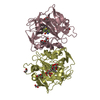
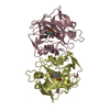
 Mass: 92.094 Da / Num. of mol.: 2 / Source method: obtained synthetically / Formula: C3H8O3
 Mass: 35.453 Da / Num. of mol.: 1 / Source method: obtained synthetically / Formula: Cl
 Mass: 18.015 Da / Num. of mol.: 454 / Source method: isolated from a natural source / Formula: H2O
 Sample preparation
 / Beamline: 8-BM / Wavelength: 0.9464, 0.979326, 0.979179
 Processing