 Movie
Movie Controller

[English] 日本語
 Yorodumi
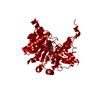
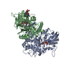
- PDB-6z5v: CRYSTAL STRUCTURE OF RAT PEROXISOMAL MULTIFUNCTIONAL ENZYME TYPE-... -
+ Open data

- Basic information
| Entry | Database: PDB / ID: 6z5v | ||||||
|---|---|---|---|---|---|---|---|
| Title | CRYSTAL STRUCTURE OF RAT PEROXISOMAL MULTIFUNCTIONAL ENZYME TYPE-1 (RPMFE1) COMPLEXED WITH 3-KETODECANOYL-COA IN CROTONASE FOLD AND OXIDISED NICOTINAMIDE ADENINE DINUCLEOTIDE IN HAD FOLD | ||||||
 Components Components | Peroxisomal bifunctional enzyme | ||||||
 Keywords Keywords | OXIDOREDUCTASE / 3-KETODECANOYL-COA / BETA-OXIDATION / PEROXISOME. | ||||||
| Function / homology |  Function and homology information Function and homology informationBeta-oxidation of very long chain fatty acids / intramolecular oxidoreductase activity, transposing C=C bonds / Peroxisomal protein import / fatty acid derivative biosynthetic process / fatty acid beta-oxidation using acyl-CoA oxidase / alpha-linolenic acid metabolic process / Delta3-Delta2-enoyl-CoA isomerase / delta(3)-delta(2)-enoyl-CoA isomerase activity / long-chain (3S)-3-hydroxyacyl-CoA dehydrogenase (NAD+) activity / 3-hydroxyacyl-CoA dehydratase activity ...Beta-oxidation of very long chain fatty acids / intramolecular oxidoreductase activity, transposing C=C bonds / Peroxisomal protein import / fatty acid derivative biosynthetic process / fatty acid beta-oxidation using acyl-CoA oxidase / alpha-linolenic acid metabolic process / Delta3-Delta2-enoyl-CoA isomerase / delta(3)-delta(2)-enoyl-CoA isomerase activity / long-chain (3S)-3-hydroxyacyl-CoA dehydrogenase (NAD+) activity / 3-hydroxyacyl-CoA dehydratase activity / 3-hydroxyacyl-CoA dehydrogenase / unsaturated fatty acid biosynthetic process / enoyl-CoA hydratase / (3S)-3-hydroxyacyl-CoA dehydrogenase (NAD+) activity / enoyl-CoA hydratase activity / long-chain fatty acid biosynthetic process / fatty acid beta-oxidation / peroxisomal matrix / NAD+ binding / peroxisome / enzyme binding / cytosol Similarity search - Function | ||||||
| Biological species |  | ||||||
| Method |  X-RAY DIFFRACTION / SYNCHROTRON / MOLECULAR REPLACEMENT / Resolution: 2.33 Å X-RAY DIFFRACTION / SYNCHROTRON / MOLECULAR REPLACEMENT / Resolution: 2.33 Å | ||||||
 Authors Authors | Wierenga, R.K. / Sridhar, S. / Kiema, T.R. | ||||||
 Citation Citation | Journal: Acta Crystallogr D Struct Biol / Year: 2020 Title: Crystallographic binding studies of rat peroxisomal multifunctional enzyme type 1 with 3-ketodecanoyl-CoA: capturing active and inactive states of its hydratase and dehydrogenase catalytic sites. Authors: Sridhar, S. / Schmitz, W. / Hiltunen, J.K. / Venkatesan, R. / Bergmann, U. / Kiema, T.R. / Wierenga, R.K. | ||||||
| History |
|
- Structure visualization
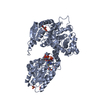
| Structure viewer | Molecule: MolmilJmol/JSmol |
|---|
- Downloads & links
-Download
| PDBx/mmCIF format | 6z5v.cif.gz | 536.4 KB | Display | PDBx/mmCIF format |
|---|---|---|---|---|
| PDB format | pdb6z5v.ent.gz | Display | PDB format | |
| PDBx/mmJSON format | 6z5v.json.gz | Tree view | PDBx/mmJSON format | |
| Others |  Other downloads Other downloads |
-Validation report
| Arichive directory | https://data.pdbj.org/pub/pdb/validation_reports/z5/6z5vftp://data.pdbj.org/pub/pdb/validation_reports/z5/6z5v | HTTPS FTP |
|---|
-Related structure data
| Related structure data |  5omoSC 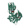 6z5fC  6z5oC S: Starting model for refinement C: citing same article ( |
|---|---|
| Similar structure data | |
| Experimental dataset #1 | Data reference: 10.23729/07397da2-8059-4c31-a9a7-9cd1b7ba6506 Data set type: diffraction image data |


-Links
 PDBj
PDBj


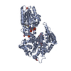
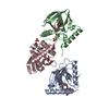
- Assembly
| Deposited unit | 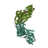
| ||||||||
|---|---|---|---|---|---|---|---|---|---|
| 1 | 
| ||||||||
| 2 | 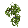
| ||||||||
| Unit cell |
|
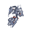
-Components
-Protein , 1 types, 2 molecules AAABBB
| #1: Protein | Mass: 80931.352 Da / Num. of mol.: 2 / Mutation: 0 Source method: isolated from a genetically manipulated source Details: Peroxisome / Source: (gene. exp.)  References: UniProt: P07896, enoyl-CoA hydratase, Delta3-Delta2-enoyl-CoA isomerase, 3-hydroxyacyl-CoA dehydrogenase |
|---|
-Non-polymers , 5 types, 176 molecules 
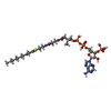


| #2: Chemical |  Mass: 663.425 Da / Num. of mol.: 2 / Source method: obtained synthetically / Formula: C21H27N7O14P2 / Feature type: SUBJECT OF INVESTIGATION / Comment: NAD*YM Mass: 663.425 Da / Num. of mol.: 2 / Source method: obtained synthetically / Formula: C21H27N7O14P2 / Feature type: SUBJECT OF INVESTIGATION / Comment: NAD*YM#3: Chemical |  Mass: 935.767 Da / Num. of mol.: 2 / Source method: obtained synthetically / Formula: C31H52N7O18P3S / Feature type: SUBJECT OF INVESTIGATION Mass: 935.767 Da / Num. of mol.: 2 / Source method: obtained synthetically / Formula: C31H52N7O18P3S / Feature type: SUBJECT OF INVESTIGATION#4: Chemical | ChemComp-GOL / |  Mass: 92.094 Da / Num. of mol.: 1 / Source method: obtained synthetically / Formula: C3H8O3 / Feature type: SUBJECT OF INVESTIGATION Mass: 92.094 Da / Num. of mol.: 1 / Source method: obtained synthetically / Formula: C3H8O3 / Feature type: SUBJECT OF INVESTIGATION#5: Chemical | ChemComp-SO4 /  Mass: 96.063 Da / Num. of mol.: 4 / Source method: obtained synthetically / Formula: SO4 / Feature type: SUBJECT OF INVESTIGATION Mass: 96.063 Da / Num. of mol.: 4 / Source method: obtained synthetically / Formula: SO4 / Feature type: SUBJECT OF INVESTIGATION#6: Water | ChemComp-HOH / | Mass: 18.015 Da / Num. of mol.: 167 / Source method: isolated from a natural source / Formula: H2O |
|---|
-Details
| Has ligand of interest | Y |
|---|
-Experimental details
-Experiment
| Experiment | Method: X-RAY DIFFRACTION / Number of used crystals: 1 |
|---|
- Sample preparation
| Crystal | Density Matthews: 2.8 Å3/Da / Density % sol: 56.6 % |
|---|---|
| Crystal grow | Temperature: 295 K / Method: vapor diffusion, sitting drop / pH: 6 Details: 125mM MES, pH 6; 17%w/v PEG4000; 175mM ammonium sulfate |
-Data collection
| Diffraction | Mean temperature: 100 K / Serial crystal experiment: N |
|---|---|
| Diffraction source | Source: SYNCHROTRON / Site: Diamond  / Beamline: I03 / Wavelength: 0.9763 Å / Beamline: I03 / Wavelength: 0.9763 Å |
| Detector | Type: DECTRIS EIGER2 X 16M / Detector: PIXEL / Date: Oct 12, 2019 |
| Radiation | Monochromator: M / Protocol: SINGLE WAVELENGTH / Monochromatic (M) / Laue (L): M / Scattering type: x-ray |
| Radiation wavelength | Wavelength: 0.9763 Å / Relative weight: 1 |
| Reflection | Resolution: 2.32→29.15 Å / Num. obs: 80007 / % possible obs: 99.2 % / Redundancy: 6.66 % / Biso Wilson estimate: 53.7 Å2 / CC1/2: 0.999 / Rmerge(I) obs: 0.053 / Rpim(I) all: 0.022 / Net I/σ(I): 17.9 |
| Reflection shell | Resolution: 2.33→2.37 Å / Rmerge(I) obs: 0.798 / Mean I/σ(I) obs: 2.1 / Num. unique obs: 3880 / CC1/2: 0.649 / Rpim(I) all: 0.359 / % possible all: 86.6 |
- Processing
| Software |
| |||||||||||||||||||||||||||||||||||||||||||||||||||||||||||||||||||||||||||||||||||||||||||||||||||||||||||||||||||||||||||||||||||||||||||||||||||||||||||||||||||||||||||||||
|---|---|---|---|---|---|---|---|---|---|---|---|---|---|---|---|---|---|---|---|---|---|---|---|---|---|---|---|---|---|---|---|---|---|---|---|---|---|---|---|---|---|---|---|---|---|---|---|---|---|---|---|---|---|---|---|---|---|---|---|---|---|---|---|---|---|---|---|---|---|---|---|---|---|---|---|---|---|---|---|---|---|---|---|---|---|---|---|---|---|---|---|---|---|---|---|---|---|---|---|---|---|---|---|---|---|---|---|---|---|---|---|---|---|---|---|---|---|---|---|---|---|---|---|---|---|---|---|---|---|---|---|---|---|---|---|---|---|---|---|---|---|---|---|---|---|---|---|---|---|---|---|---|---|---|---|---|---|---|---|---|---|---|---|---|---|---|---|---|---|---|---|---|---|---|---|---|
| Refinement | Method to determine structure: MOLECULAR REPLACEMENT Starting model: 5OMO Resolution: 2.33→29.149 Å / Cor.coef. Fo:Fc: 0.96 / Cor.coef. Fo:Fc free: 0.952 / Cross valid method: FREE R-VALUE / ESU R: 0.298 / ESU R Free: 0.208 Details: Hydrogens have been added in their riding positions
| |||||||||||||||||||||||||||||||||||||||||||||||||||||||||||||||||||||||||||||||||||||||||||||||||||||||||||||||||||||||||||||||||||||||||||||||||||||||||||||||||||||||||||||||
| Solvent computation | Ion probe radii: 0.8 Å / Shrinkage radii: 0.8 Å / VDW probe radii: 1.2 Å / Solvent model: MASK BULK SOLVENT | |||||||||||||||||||||||||||||||||||||||||||||||||||||||||||||||||||||||||||||||||||||||||||||||||||||||||||||||||||||||||||||||||||||||||||||||||||||||||||||||||||||||||||||||
| Displacement parameters | Biso mean: 70 Å2
| |||||||||||||||||||||||||||||||||||||||||||||||||||||||||||||||||||||||||||||||||||||||||||||||||||||||||||||||||||||||||||||||||||||||||||||||||||||||||||||||||||||||||||||||
| Refinement step | Cycle: LAST / Resolution: 2.33→29.149 Å
| |||||||||||||||||||||||||||||||||||||||||||||||||||||||||||||||||||||||||||||||||||||||||||||||||||||||||||||||||||||||||||||||||||||||||||||||||||||||||||||||||||||||||||||||
| Refine LS restraints |
| |||||||||||||||||||||||||||||||||||||||||||||||||||||||||||||||||||||||||||||||||||||||||||||||||||||||||||||||||||||||||||||||||||||||||||||||||||||||||||||||||||||||||||||||
| LS refinement shell |
| |||||||||||||||||||||||||||||||||||||||||||||||||||||||||||||||||||||||||||||||||||||||||||||||||||||||||||||||||||||||||||||||||||||||||||||||||||||||||||||||||||||||||||||||
| Refinement TLS params. | Method: refined / Refine-ID: X-RAY DIFFRACTION
| |||||||||||||||||||||||||||||||||||||||||||||||||||||||||||||||||||||||||||||||||||||||||||||||||||||||||||||||||||||||||||||||||||||||||||||||||||||||||||||||||||||||||||||||
| Refinement TLS group |
|
