 Movie
Movie Controller

[English] 日本語
 Yorodumi
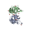
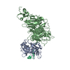
- PDB-5omo: CRYSTAL STRUCTURE OF RAT PEROXISOMAL MULTIFUNCTIONAL ENZYME TYPE-... -
+ Open data

- Basic information
| Entry | Database: PDB / ID: 5omo | |||||||||
|---|---|---|---|---|---|---|---|---|---|---|
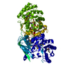
| Title | CRYSTAL STRUCTURE OF RAT PEROXISOMAL MULTIFUNCTIONAL ENZYME TYPE-1 (RPMFE1) COMPLEXED WITH WITH 3S-HYDROXY-DECANOYL-COA AND 3-KETO-DECANOYL-COA | |||||||||
 Components Components | Peroxisomal bifunctional enzyme | |||||||||
 Keywords Keywords | OXIDOREDUCTASE / 3S-HYDROXY-DECANOYL-COA / 3-KETO-DECANOYL-COA / MFE1 / BETA-OXIDATION / FATTY ACID / CROTONASE / 3-HYDROXYACYL-COA- DEHYDROGENASE / HYDRATASE | |||||||||
| Function / homology |  Function and homology information Function and homology informationBeta-oxidation of very long chain fatty acids / intramolecular oxidoreductase activity, transposing C=C bonds / Peroxisomal protein import / fatty acid beta-oxidation using acyl-CoA oxidase / fatty acid derivative biosynthetic process / alpha-linolenic acid metabolic process / Delta3-Delta2-enoyl-CoA isomerase / delta(3)-delta(2)-enoyl-CoA isomerase activity / long-chain (3S)-3-hydroxyacyl-CoA dehydrogenase (NAD+) activity / 3-hydroxyacyl-CoA dehydratase activity ...Beta-oxidation of very long chain fatty acids / intramolecular oxidoreductase activity, transposing C=C bonds / Peroxisomal protein import / fatty acid beta-oxidation using acyl-CoA oxidase / fatty acid derivative biosynthetic process / alpha-linolenic acid metabolic process / Delta3-Delta2-enoyl-CoA isomerase / delta(3)-delta(2)-enoyl-CoA isomerase activity / long-chain (3S)-3-hydroxyacyl-CoA dehydrogenase (NAD+) activity / 3-hydroxyacyl-CoA dehydratase activity / 3-hydroxyacyl-CoA dehydrogenase / unsaturated fatty acid biosynthetic process / enoyl-CoA hydratase / (3S)-3-hydroxyacyl-CoA dehydrogenase (NAD+) activity / enoyl-CoA hydratase activity / long-chain fatty acid biosynthetic process / fatty acid beta-oxidation / peroxisomal matrix / NAD+ binding / peroxisome / enzyme binding / cytosol Similarity search - Function | |||||||||
| Biological species |  | |||||||||
| Method |  X-RAY DIFFRACTION / SYNCHROTRON / FOURIER SYNTHESIS / Resolution: 2.49 Å X-RAY DIFFRACTION / SYNCHROTRON / FOURIER SYNTHESIS / Resolution: 2.49 Å | |||||||||
 Authors Authors | Kasaragod, P. / Kiema, T.-R. / Schmitz, W. / Hiltunen, J.K. / Wierenga, R.K. | |||||||||
 Citation Citation | Journal: Acta Crystallogr D Struct Biol / Year: 2020 Title: Crystallographic binding studies of rat peroxisomal multifunctional enzyme type 1 with 3-ketodecanoyl-CoA: capturing active and inactive states of its hydratase and dehydrogenase catalytic sites. Authors: Sridhar, S. / Schmitz, W. / Hiltunen, J.K. / Venkatesan, R. / Bergmann, U. / Kiema, T.R. / Wierenga, R.K. | |||||||||
| History |
|
- Structure visualization
| Structure viewer | Molecule: MolmilJmol/JSmol |
|---|
- Downloads & links
-Download
| PDBx/mmCIF format | 5omo.cif.gz | 571.1 KB | Display | PDBx/mmCIF format |
|---|---|---|---|---|
| PDB format | pdb5omo.ent.gz | 469.5 KB | Display | PDB format |
| PDBx/mmJSON format | 5omo.json.gz | Tree view | PDBx/mmJSON format | |
| Others |  Other downloads Other downloads |
-Validation report
| Arichive directory | https://data.pdbj.org/pub/pdb/validation_reports/om/5omoftp://data.pdbj.org/pub/pdb/validation_reports/om/5omo | HTTPS FTP |
|---|
-Related structure data
| Related structure data | 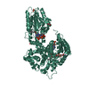 6z5fC  6z5oC 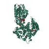 6z5vC  2x58S S: Starting model for refinement C: citing same article ( |
|---|---|
| Similar structure data |


-Links
 PDBj
PDBj


- Assembly
| Deposited unit | 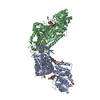
| ||||||||||||||||||
|---|---|---|---|---|---|---|---|---|---|---|---|---|---|---|---|---|---|---|---|
| 1 | 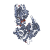
| ||||||||||||||||||
| 2 | 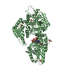
| ||||||||||||||||||
| Unit cell |
| ||||||||||||||||||
| Noncrystallographic symmetry (NCS) | NCS domain:
NCS domain segments: Component-ID: _ / Ens-ID: 1 / Beg auth comp-ID: HIS / Beg label comp-ID: HIS / End auth comp-ID: GLY / End label comp-ID: GLY / Refine code: _ / Auth seq-ID: 0 - 716 / Label seq-ID: 20 - 736
|
-Components
-Protein , 1 types, 2 molecules AB
| #1: Protein | Mass: 80931.352 Da / Num. of mol.: 2 Source method: isolated from a genetically manipulated source Details: 3S-HYDROXY-DECANOYL-COA BOUND IN THE CROTONASE DOMAIN AND 3-KETO-DECANOYL-COA BOUND IN THE 3-HYDROXYACYL-COA-DEHYDROGENASE DOMAIN Source: (gene. exp.)  References: UniProt: P07896, enoyl-CoA hydratase, Delta3-Delta2-enoyl-CoA isomerase, 3-hydroxyacyl-CoA dehydrogenase |
|---|
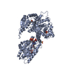
-Non-polymers , 5 types, 294 molecules 


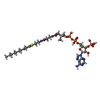

| #2: Chemical | ChemComp-SO4 /  Mass: 96.063 Da / Num. of mol.: 6 / Source method: obtained synthetically / Formula: SO4 Mass: 96.063 Da / Num. of mol.: 6 / Source method: obtained synthetically / Formula: SO4#3: Chemical |  Mass: 92.094 Da / Num. of mol.: 3 / Source method: obtained synthetically / Formula: C3H8O3 Mass: 92.094 Da / Num. of mol.: 3 / Source method: obtained synthetically / Formula: C3H8O3#4: Chemical |  Mass: 933.751 Da / Num. of mol.: 2 / Source method: obtained synthetically / Formula: C31H50N7O18P3S / Feature type: SUBJECT OF INVESTIGATION Mass: 933.751 Da / Num. of mol.: 2 / Source method: obtained synthetically / Formula: C31H50N7O18P3S / Feature type: SUBJECT OF INVESTIGATION#5: Chemical | ChemComp-ZOZ / |  Mass: 935.767 Da / Num. of mol.: 1 / Source method: obtained synthetically / Formula: C31H52N7O18P3S / Feature type: SUBJECT OF INVESTIGATION Mass: 935.767 Da / Num. of mol.: 1 / Source method: obtained synthetically / Formula: C31H52N7O18P3S / Feature type: SUBJECT OF INVESTIGATION#6: Water | ChemComp-HOH / | Mass: 18.015 Da / Num. of mol.: 282 / Source method: isolated from a natural source / Formula: H2O |
|---|
-Details
| Has protein modification | N |
|---|
-Experimental details
-Experiment
| Experiment | Method: X-RAY DIFFRACTION / Number of used crystals: 1 |
|---|
- Sample preparation
| Crystal | Density Matthews: 2.81 Å3/Da / Density % sol: 56.24 % |
|---|---|
| Crystal grow | Temperature: 295 K / Method: vapor diffusion / pH: 6 Details: 100MM MES PH 6.0, 150MM AMMONIUM SULPHATE, 15% W/V PEG4000 |
-Data collection
| Diffraction | Mean temperature: 100 K |
|---|---|
| Diffraction source | Source: SYNCHROTRON / Site: ESRF  / Beamline: ID14-4 / Wavelength: 0.93928 Å / Beamline: ID14-4 / Wavelength: 0.93928 Å |
| Detector | Type: ADSC QUANTUM 315r / Detector: CCD / Date: Nov 29, 2010 |
| Radiation | Protocol: SINGLE WAVELENGTH / Monochromatic (M) / Laue (L): M / Scattering type: x-ray |
| Radiation wavelength | Wavelength: 0.93928 Å / Relative weight: 1 |
| Reflection | Resolution: 2.49→47.9 Å / Num. obs: 64401 / % possible obs: 99.2 % / Redundancy: 4.4 % / Rmerge(I) obs: 0.06 / Rpim(I) all: 0.029 / Net I/σ(I): 16.6 |
| Reflection shell | Resolution: 2.49→2.62 Å / Redundancy: 4 % / Rmerge(I) obs: 0.28 / Mean I/σ(I) obs: 4.2 / Num. unique obs: 8976 / Rpim(I) all: 0.158 / % possible all: 96.1 |
- Processing
| Software |
| ||||||||||||||||||||||||||||||||||||||||||||||||||||||||||||||||||||||||||||||||||||||||||||||||||||||||||||||||||||||||||||||||||||||||||||||||||||||||||||||||||||||||||||||||||||||||||||||||
|---|---|---|---|---|---|---|---|---|---|---|---|---|---|---|---|---|---|---|---|---|---|---|---|---|---|---|---|---|---|---|---|---|---|---|---|---|---|---|---|---|---|---|---|---|---|---|---|---|---|---|---|---|---|---|---|---|---|---|---|---|---|---|---|---|---|---|---|---|---|---|---|---|---|---|---|---|---|---|---|---|---|---|---|---|---|---|---|---|---|---|---|---|---|---|---|---|---|---|---|---|---|---|---|---|---|---|---|---|---|---|---|---|---|---|---|---|---|---|---|---|---|---|---|---|---|---|---|---|---|---|---|---|---|---|---|---|---|---|---|---|---|---|---|---|---|---|---|---|---|---|---|---|---|---|---|---|---|---|---|---|---|---|---|---|---|---|---|---|---|---|---|---|---|---|---|---|---|---|---|---|---|---|---|---|---|---|---|---|---|---|---|---|---|
| Refinement | Method to determine structure: FOURIER SYNTHESIS Starting model: 2X58 Resolution: 2.49→47.9 Å / Cor.coef. Fo:Fc: 0.944 / Cor.coef. Fo:Fc free: 0.921 / SU B: 16.867 / SU ML: 0.189 / Cross valid method: THROUGHOUT / ESU R: 0.436 / ESU R Free: 0.253 / Details: HYDROGENS HAVE BEEN ADDED IN THE RIDING POSITIONS
| ||||||||||||||||||||||||||||||||||||||||||||||||||||||||||||||||||||||||||||||||||||||||||||||||||||||||||||||||||||||||||||||||||||||||||||||||||||||||||||||||||||||||||||||||||||||||||||||||
| Solvent computation | Ion probe radii: 0.8 Å / Shrinkage radii: 0.8 Å / VDW probe radii: 1.2 Å | ||||||||||||||||||||||||||||||||||||||||||||||||||||||||||||||||||||||||||||||||||||||||||||||||||||||||||||||||||||||||||||||||||||||||||||||||||||||||||||||||||||||||||||||||||||||||||||||||
| Displacement parameters | Biso mean: 53.337 Å2
| ||||||||||||||||||||||||||||||||||||||||||||||||||||||||||||||||||||||||||||||||||||||||||||||||||||||||||||||||||||||||||||||||||||||||||||||||||||||||||||||||||||||||||||||||||||||||||||||||
| Refinement step | Cycle: 1 / Resolution: 2.49→47.9 Å
| ||||||||||||||||||||||||||||||||||||||||||||||||||||||||||||||||||||||||||||||||||||||||||||||||||||||||||||||||||||||||||||||||||||||||||||||||||||||||||||||||||||||||||||||||||||||||||||||||
| Refine LS restraints |
| ||||||||||||||||||||||||||||||||||||||||||||||||||||||||||||||||||||||||||||||||||||||||||||||||||||||||||||||||||||||||||||||||||||||||||||||||||||||||||||||||||||||||||||||||||||||||||||||||
| Refine LS restraints NCS | Ens-ID: 1 / Number: 44904 / Refine-ID: X-RAY DIFFRACTION / Type: interatomic distance / Rms dev position: 0.09 Å / Weight position: 0.05
| ||||||||||||||||||||||||||||||||||||||||||||||||||||||||||||||||||||||||||||||||||||||||||||||||||||||||||||||||||||||||||||||||||||||||||||||||||||||||||||||||||||||||||||||||||||||||||||||||
| LS refinement shell | Resolution: 2.487→2.552 Å / Total num. of bins used: 20
| ||||||||||||||||||||||||||||||||||||||||||||||||||||||||||||||||||||||||||||||||||||||||||||||||||||||||||||||||||||||||||||||||||||||||||||||||||||||||||||||||||||||||||||||||||||||||||||||||
| Refinement TLS params. | Method: refined / Refine-ID: X-RAY DIFFRACTION
| ||||||||||||||||||||||||||||||||||||||||||||||||||||||||||||||||||||||||||||||||||||||||||||||||||||||||||||||||||||||||||||||||||||||||||||||||||||||||||||||||||||||||||||||||||||||||||||||||
| Refinement TLS group |
|
