 Movie
Movie Controller


+ Open data

- Basic information
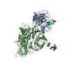
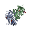
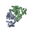
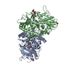
| Entry | Database: PDB / ID: 4o3u | ||||||
|---|---|---|---|---|---|---|---|
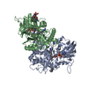
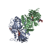
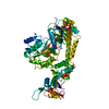
| Title | Zymogen HGF-beta/MET with Zymogen Activator Peptide ZAP2.3 | ||||||
 Components Components |
| ||||||
 Keywords Keywords | transferase/growth factor / trypsin homoloy / receptor activation / transferase-growth factor complex | ||||||
| Function / homology |  Function and homology information Function and homology informationregulation of branching involved in salivary gland morphogenesis by mesenchymal-epithelial signaling / skeletal muscle cell proliferation / regulation of p38MAPK cascade / hepatocyte growth factor receptor activity / Drug-mediated inhibition of MET activation / MET activates STAT3 / negative regulation of hydrogen peroxide-mediated programmed cell death / MET Receptor Activation / MET interacts with TNS proteins / endothelial cell morphogenesis ...regulation of branching involved in salivary gland morphogenesis by mesenchymal-epithelial signaling / skeletal muscle cell proliferation / regulation of p38MAPK cascade / hepatocyte growth factor receptor activity / Drug-mediated inhibition of MET activation / MET activates STAT3 / negative regulation of hydrogen peroxide-mediated programmed cell death / MET Receptor Activation / MET interacts with TNS proteins / endothelial cell morphogenesis / semaphorin receptor activity / MET receptor recycling / pancreas development / MET activates PTPN11 / hepatocyte growth factor receptor signaling pathway / MET activates RAP1 and RAC1 / cellular response to hepatocyte growth factor stimulus / Sema4D mediated inhibition of cell attachment and migration / positive regulation of endothelial cell chemotaxis / MET activates PI3K/AKT signaling / MET activates PTK2 signaling / positive regulation of DNA biosynthetic process / branching morphogenesis of an epithelial tube / negative regulation of release of cytochrome c from mitochondria / positive chemotaxis / chemoattractant activity / negative regulation of interleukin-6 production / semaphorin-plexin signaling pathway / myoblast proliferation / positive regulation of interleukin-10 production / epithelial to mesenchymal transition / positive regulation of osteoblast differentiation / Regulation of MITF-M-dependent genes involved in cell cycle and proliferation / negative regulation of extrinsic apoptotic signaling pathway via death domain receptors / MET activates RAS signaling / epithelial cell proliferation / MECP2 regulates neuronal receptors and channels / platelet alpha granule lumen / Interleukin-7 signaling / cell surface receptor protein tyrosine kinase signaling pathway / basal plasma membrane / molecular function activator activity / excitatory postsynaptic potential / liver development / negative regulation of autophagy / InlB-mediated entry of Listeria monocytogenes into host cell / growth factor activity / cell chemotaxis / receptor protein-tyrosine kinase / Negative regulation of MET activity / negative regulation of inflammatory response / cell morphogenesis / neuron differentiation / Constitutive Signaling by Aberrant PI3K in Cancer / Platelet degranulation / PIP3 activates AKT signaling / mitotic cell cycle / PI5P, PP2A and IER3 Regulate PI3K/AKT Signaling / RAF/MAP kinase cascade / protein tyrosine kinase activity / Interleukin-4 and Interleukin-13 signaling / protein phosphatase binding / positive regulation of MAPK cascade / positive regulation of phosphatidylinositol 3-kinase/protein kinase B signal transduction / cell surface receptor signaling pathway / postsynapse / signaling receptor complex / positive regulation of cell migration / signaling receptor binding / negative regulation of apoptotic process / cell surface / positive regulation of transcription by RNA polymerase II / : / extracellular region / ATP binding / membrane / identical protein binding / plasma membrane Similarity search - Function | ||||||
| Biological species |  Homo sapiens (human) Homo sapiens (human)synthetic (others) | ||||||
| Method |  X-RAY DIFFRACTION / SYNCHROTRON / MOLECULAR REPLACEMENT / Resolution: 3.04 Å X-RAY DIFFRACTION / SYNCHROTRON / MOLECULAR REPLACEMENT / Resolution: 3.04 Å | ||||||
 Authors Authors | Eigenbrot, C. / Landgraf, K.E. / Steffek, M. | ||||||
 Citation Citation | Journal: Nat.Chem.Biol. / Year: 2014 Title: An allosteric switch for pro-HGF/Met signaling using zymogen activator peptides. Authors: Landgraf, K.E. / Steffek, M. / Quan, C. / Tom, J. / Yu, C. / Santell, L. / Maun, H.R. / Eigenbrot, C. / Lazarus, R.A. | ||||||
| History |
|
- Structure visualization
| Structure viewer | Molecule: MolmilJmol/JSmol |
|---|
- Downloads & links
-Download
| PDBx/mmCIF format | 4o3u.cif.gz | 159 KB | Display | PDBx/mmCIF format |
|---|---|---|---|---|
| PDB format | pdb4o3u.ent.gz | 122.8 KB | Display | PDB format |
| PDBx/mmJSON format | 4o3u.json.gz | Tree view | PDBx/mmJSON format | |
| Others |  Other downloads Other downloads |
-Validation report
| Arichive directory | https://data.pdbj.org/pub/pdb/validation_reports/o3/4o3uftp://data.pdbj.org/pub/pdb/validation_reports/o3/4o3u | HTTPS FTP |
|---|
-Related structure data
| Related structure data |  4o3tC 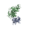 1shyS S: Starting model for refinement C: citing same article ( |
|---|---|
| Similar structure data |


-Links
 PDBj
PDBj


- Assembly
| Deposited unit | 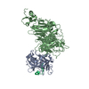
| ||||||||
|---|---|---|---|---|---|---|---|---|---|
| 1 |
| ||||||||
| Unit cell |
|
-Components
| #1: Protein | Mass: 26800.920 Da / Num. of mol.: 1 / Fragment: HGF-beta (UNP Residues 25-567) / Mutation: V495G/C604S Source method: isolated from a genetically manipulated source Source: (gene. exp.) Homo sapiens (human) / Gene: HGF, HPTA / Production host:   Spodoptera frugiperda (fall armyworm) / References: UniProt: P14210 Spodoptera frugiperda (fall armyworm) / References: UniProt: P14210 |
|---|---|
| #2: Protein | Mass: 62714.141 Da / Num. of mol.: 1 / Fragment: Sema-PSI (UNP Residues 496-728) / Mutation: L303K/V304R/P305K/R306K/G307R Source method: isolated from a genetically manipulated source Source: (gene. exp.) Homo sapiens (human) / Gene: MET / Production host: Spodoptera frugiperda (fall armyworm)References: UniProt: P08581, receptor protein-tyrosine kinase |
| #3: Protein/peptide | Mass: 1786.081 Da / Num. of mol.: 1 / Source method: obtained synthetically / Details: synthetic peptide library displayed on phage / Source: (synth.) synthetic (others) |
| Has protein modification | Y |
-Experimental details
-Experiment
| Experiment | Method: X-RAY DIFFRACTION / Number of used crystals: 1 |
|---|
- Sample preparation
| Crystal | Density Matthews: 3.44 Å3/Da / Density % sol: 64.26 % |
|---|---|
| Crystal grow | Temperature: 293 K / Method: vapor diffusion, sitting drop / pH: 7.2 Details: 8% PEG8000, pH 7.2, VAPOR DIFFUSION, SITTING DROP, temperature 293K |
-Data collection
| Diffraction | Mean temperature: 110 K |
|---|---|
| Diffraction source | Source: SYNCHROTRON / Site: SSRL  / Beamline: BL12-2 / Wavelength: 0.9794 Å / Beamline: BL12-2 / Wavelength: 0.9794 Å |
| Detector | Type: PSI PILATUS 6M / Detector: PIXEL / Date: May 23, 2012 |
| Radiation | Monochromator: Si(111) / Protocol: SINGLE WAVELENGTH / Monochromatic (M) / Laue (L): M / Scattering type: x-ray |
| Radiation wavelength | Wavelength: 0.9794 Å / Relative weight: 1 |
| Reflection | Resolution: 3.04→34.76 Å / Num. all: 24419 / Num. obs: 24340 / % possible obs: 97.5 % / Observed criterion σ(F): 0 / Observed criterion σ(I): -3 / Redundancy: 6.6 % / Biso Wilson estimate: 102.77 Å2 / Rsym value: 0.056 / Net I/σ(I): 23 |
- Processing
| Software |
| ||||||||||||||||||||||||||||||||||||||||||||||||||||||||||||||||||
|---|---|---|---|---|---|---|---|---|---|---|---|---|---|---|---|---|---|---|---|---|---|---|---|---|---|---|---|---|---|---|---|---|---|---|---|---|---|---|---|---|---|---|---|---|---|---|---|---|---|---|---|---|---|---|---|---|---|---|---|---|---|---|---|---|---|---|---|
| Refinement | Method to determine structure: MOLECULAR REPLACEMENT Starting model: PDB ENTRY 1SHY Resolution: 3.04→34.76 Å / Cor.coef. Fo:Fc: 0.8969 / Cor.coef. Fo:Fc free: 0.8574 / SU R Cruickshank DPI: 1.39 / Isotropic thermal model: individual atom isotropic / Cross valid method: THROUGHOUT / σ(F): 0 / Stereochemistry target values: Engh & Huber
| ||||||||||||||||||||||||||||||||||||||||||||||||||||||||||||||||||
| Displacement parameters | Biso mean: 94.12 Å2
| ||||||||||||||||||||||||||||||||||||||||||||||||||||||||||||||||||
| Refine analyze | Luzzati coordinate error obs: 0.593 Å | ||||||||||||||||||||||||||||||||||||||||||||||||||||||||||||||||||
| Refinement step | Cycle: LAST / Resolution: 3.04→34.76 Å
| ||||||||||||||||||||||||||||||||||||||||||||||||||||||||||||||||||
| Refine LS restraints |
| ||||||||||||||||||||||||||||||||||||||||||||||||||||||||||||||||||
| LS refinement shell | Resolution: 3.04→3.17 Å / Total num. of bins used: 12
|
