multimeric ribonuclease P complex / nucleolar ribonuclease P complex / ribonuclease P RNA binding / ribonuclease MRP complex / tRNA processing in the nucleus / ribonuclease P activity / tRNA 5'-leader removal / Major pathway of rRNA processing in the nucleolus and cytosol / endonucleolytic cleavage in ITS1 to separate SSU-rRNA from 5.8S rRNA and LSU-rRNA from tricistronic rRNA transcript (SSU-rRNA, 5.8S rRNA, LSU-rRNA) / nucleoplasm / nucleus Similarity search - Function
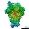
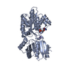
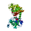
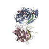
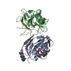
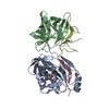
Journal: Cell / Year: 2018 Title: Cryo-EM Structure of the Human Ribonuclease P Holoenzyme. Authors: Jian Wu / Shuangshuang Niu / Ming Tan / Chenhui Huang / Mingyue Li / Yang Song / Qianmin Wang / Juan Chen / Shaohua Shi / Pengfei Lan / Ming Lei / Abstract: Ribonuclease (RNase) P is a ubiquitous ribozyme that cleaves the 5' leader from precursor tRNAs. Here, we report cryo-electron microscopy structures of the human nuclear RNase P alone and in ...Ribonuclease (RNase) P is a ubiquitous ribozyme that cleaves the 5' leader from precursor tRNAs. Here, we report cryo-electron microscopy structures of the human nuclear RNase P alone and in complex with tRNA. Human RNase P is a large ribonucleoprotein complex that contains 10 protein components and one catalytic RNA. The protein components form an interlocked clamp that stabilizes the RNA in a conformation optimal for substrate binding. Human RNase P recognizes the tRNA using a double-anchor mechanism through both protein-RNA and RNA-RNA interactions. Structural comparison of the apo and tRNA-bound human RNase P reveals that binding of tRNA induces a local conformational change in the catalytic center, transforming the ribozyme into an active state. Our results also provide an evolutionary model depicting how auxiliary RNA elements in bacterial RNase P, essential for substrate binding, and catalysis, were replaced by the much more complex and multifunctional protein components in higher organisms.
Method to determine structure: SAD / Resolution: 2.6→43.434 Å / SU ML: 0.41 / Cross valid method: THROUGHOUT / σ(F): 1.36 / Phase error: 27.39 / Stereochemistry target values: ML
Rfactor
Num. reflection
% reflection
Rfree
0.2531
877
5.15 %
Rwork
0.2179
16156
-
obs
0.2198
17033
97.05 %
Solvent computation
Shrinkage radii: 0.9 Å / VDW probe radii: 1.11 Å / Solvent model: FLAT BULK SOLVENT MODEL
In the structure databanks used in Yorodumi, some data are registered as the other names, "COVID-19 virus" and "2019-nCoV". Here are the details of the virus and the list of structure data.
Jan 31, 2019. EMDB accession codes are about to change! (news from PDBe EMDB page)
EMDB accession codes are about to change! (news from PDBe EMDB page)
The allocation of 4 digits for EMDB accession codes will soon come to an end. Whilst these codes will remain in use, new EMDB accession codes will include an additional digit and will expand incrementally as the available range of codes is exhausted. The current 4-digit format prefixed with “EMD-” (i.e. EMD-XXXX) will advance to a 5-digit format (i.e. EMD-XXXXX), and so on. It is currently estimated that the 4-digit codes will be depleted around Spring 2019, at which point the 5-digit format will come into force.
The EM Navigator/Yorodumi systems omit the EMD- prefix.
Related info.:Q: What is EMD? / ID/Accession-code notation in Yorodumi/EM Navigator
Yorodumi is a browser for structure data from EMDB, PDB, SASBDB, etc.
This page is also the successor to EM Navigator detail page, and also detail information page/front-end page for Omokage search.
The word "yorodu" (or yorozu) is an old Japanese word meaning "ten thousand". "mi" (miru) is to see.
Related info.:EMDB / PDB / SASBDB / Comparison of 3 databanks / Yorodumi Search / Aug 31, 2016. New EM Navigator & Yorodumi / Yorodumi Papers / Jmol/JSmol / Function and homology information / Changes in new EM Navigator and Yorodumi
 Movie
Movie Controller


 Open data
 Basic information
 Components
 Keywords
 Function and homology information
 Homo sapiens (human)
 X-RAY DIFFRACTION /
  Authors
 Citation
 Structure visualization
 Downloads & links
 Other downloads
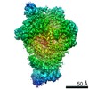


 PDBj
PDBj


 Assembly
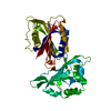


 Mass: 18.015 Da / Num. of mol.: 26 / Source method: isolated from a natural source / Formula: H2O
 Sample preparation
 Processing