 Movie
Movie Controller


+ Open data

- Basic information
| Entry | Database: PDB / ID: 6ahr | ||||||
|---|---|---|---|---|---|---|---|
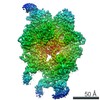
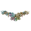
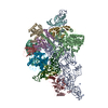
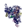
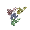
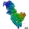
| Title | Cryo-EM structure of human Ribonuclease P | ||||||
 Components Components |
| ||||||
 Keywords Keywords | HYDROLASE/RNA / Ribonuclease P / RNA-protein complex / HYDROLASE-RNA complex | ||||||
| Function / homology |  Function and homology information Function and homology informationmultimeric ribonuclease P complex / nucleolar ribonuclease P complex / ribonuclease P RNA binding / ribonuclease MRP complex / ribonuclease P complex / tRNA decay / tRNA processing in the nucleus / ribonuclease P activity / tRNA 5'-leader removal / tRNA processing ...multimeric ribonuclease P complex / nucleolar ribonuclease P complex / ribonuclease P RNA binding / ribonuclease MRP complex / ribonuclease P complex / tRNA decay / tRNA processing in the nucleus / ribonuclease P activity / tRNA 5'-leader removal / tRNA processing / Major pathway of rRNA processing in the nucleolus and cytosol / endonucleolytic cleavage in ITS1 to separate SSU-rRNA from 5.8S rRNA and LSU-rRNA from tricistronic rRNA transcript (SSU-rRNA, 5.8S rRNA, LSU-rRNA) / fibrillar center / centriolar satellite / rRNA processing / response to xenobiotic stimulus / nucleolus / : / RNA binding / nucleoplasm / metal ion binding / nucleus / cytoplasm Similarity search - Function | ||||||
| Biological species |  Homo sapiens (human) Homo sapiens (human) | ||||||
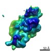
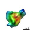
| Method | ELECTRON MICROSCOPY / single particle reconstruction / cryo EM / Resolution: 3.92 Å | ||||||
 Authors Authors | Wu, J. / Niu, S. / Tan, M. / Lan, P. / Lei, M. | ||||||
 Citation Citation | Journal: Cell / Year: 2018 Title: Cryo-EM Structure of the Human Ribonuclease P Holoenzyme. Authors: Jian Wu / Shuangshuang Niu / Ming Tan / Chenhui Huang / Mingyue Li / Yang Song / Qianmin Wang / Juan Chen / Shaohua Shi / Pengfei Lan / Ming Lei /  Abstract: Ribonuclease (RNase) P is a ubiquitous ribozyme that cleaves the 5' leader from precursor tRNAs. Here, we report cryo-electron microscopy structures of the human nuclear RNase P alone and in ...Ribonuclease (RNase) P is a ubiquitous ribozyme that cleaves the 5' leader from precursor tRNAs. Here, we report cryo-electron microscopy structures of the human nuclear RNase P alone and in complex with tRNA. Human RNase P is a large ribonucleoprotein complex that contains 10 protein components and one catalytic RNA. The protein components form an interlocked clamp that stabilizes the RNA in a conformation optimal for substrate binding. Human RNase P recognizes the tRNA using a double-anchor mechanism through both protein-RNA and RNA-RNA interactions. Structural comparison of the apo and tRNA-bound human RNase P reveals that binding of tRNA induces a local conformational change in the catalytic center, transforming the ribozyme into an active state. Our results also provide an evolutionary model depicting how auxiliary RNA elements in bacterial RNase P, essential for substrate binding, and catalysis, were replaced by the much more complex and multifunctional protein components in higher organisms. | ||||||
| History |
|
- Structure visualization
| Movie |
Movie viewer |
|---|---|
| Structure viewer | Molecule: MolmilJmol/JSmol |
- Downloads & links
-Download
| PDBx/mmCIF format | 6ahr.cif.gz | 635.5 KB | Display | PDBx/mmCIF format |
|---|---|---|---|---|
| PDB format | pdb6ahr.ent.gz | 499.8 KB | Display | PDB format |
| PDBx/mmJSON format | 6ahr.json.gz | Tree view | PDBx/mmJSON format | |
| Others |  Other downloads Other downloads |
-Validation report
| Arichive directory | https://data.pdbj.org/pub/pdb/validation_reports/ah/6ahrftp://data.pdbj.org/pub/pdb/validation_reports/ah/6ahr | HTTPS FTP |
|---|
-Related structure data
| Related structure data | 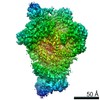 9626MC 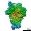 9627C 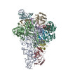 6ahuC 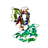 6ahvC M: map data used to model this data C: citing same article ( |
|---|---|
| Similar structure data |

-Links
 PDBj
PDBj


- Assembly
| Deposited unit | 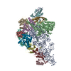
|
|---|---|
| 1 |
|
-Components
-Protein , 2 types, 2 molecules BE
| #2: Protein | Mass: 114896.141 Da / Num. of mol.: 1 / Source method: isolated from a natural source / Details: cell / Source: (natural) Homo sapiens (human) / References: UniProt: Q99575, ribonuclease P |
|---|---|
| #5: Protein | Mass: 18844.672 Da / Num. of mol.: 1 / Source method: isolated from a natural source / Details: cell / Source: (natural) Homo sapiens (human) / References: UniProt: Q969H6, ribonuclease P |
-Ribonuclease P protein subunit ... , 8 types, 9 molecules CDFGHIJKL
| #3: Protein | Mass: 31891.443 Da / Num. of mol.: 1 / Source method: isolated from a natural source / Details: cell / Source: (natural) Homo sapiens (human) / References: UniProt: P78345, ribonuclease P | ||||
|---|---|---|---|---|---|
| #4: Protein | Mass: 25474.854 Da / Num. of mol.: 1 / Source method: isolated from a natural source / Details: cell / Source: (natural) Homo sapiens (human) / References: UniProt: O95707, ribonuclease P | ||||
| #6: Protein | Mass: 20659.406 Da / Num. of mol.: 1 / Source method: isolated from a natural source / Details: cell / Source: (natural) Homo sapiens (human) / References: UniProt: Q9BUL9, ribonuclease P | ||||
| #7: Protein | Mass: 15672.866 Da / Num. of mol.: 1 / Source method: isolated from a natural source / Details: cell / Source: (natural) Homo sapiens (human) / References: UniProt: O75817, ribonuclease P | ||||
| #8: Protein | Mass: 13707.014 Da / Num. of mol.: 1 / Source method: isolated from a natural source / Details: cell / Source: (natural) Homo sapiens (human) / References: UniProt: O95059, ribonuclease P | ||||
| #9: Protein | Mass: 29364.277 Da / Num. of mol.: 2 / Source method: isolated from a natural source / Details: cell / Source: (natural) Homo sapiens (human) / References: UniProt: P78346, ribonuclease P#10: Protein | | Mass: 17596.068 Da / Num. of mol.: 1 / Source method: isolated from a natural source / Details: cell / Source: (natural) Homo sapiens (human) / References: UniProt: Q9H633, ribonuclease P#11: Protein | | Mass: 41884.844 Da / Num. of mol.: 1 / Source method: isolated from a natural source / Details: cell / Source: (natural) Homo sapiens (human) / References: UniProt: O75818, ribonuclease P |
-RNA chain / Non-polymers , 2 types, 2 molecules A
| #12: Chemical | ChemComp-ZN /  Mass: 65.409 Da / Num. of mol.: 1 / Source method: obtained synthetically / Formula: Zn Mass: 65.409 Da / Num. of mol.: 1 / Source method: obtained synthetically / Formula: Zn |
|---|---|
| #1: RNA chain | Mass: 110244.172 Da / Num. of mol.: 1 / Source method: isolated from a natural source / Details: cell / Source: (natural) Homo sapiens (human) / References: GenBank: 31969 |
-Experimental details
-Experiment
| Experiment | Method: ELECTRON MICROSCOPY |
|---|---|
| EM experiment | Aggregation state: PARTICLE / 3D reconstruction method: single particle reconstruction |
- Sample preparation
| Component | Name: RNase P / Type: COMPLEX / Entity ID: #1-#11 / Source: NATURAL |
|---|---|
| Source (natural) | Organism: Homo sapiens (human) |
| Buffer solution | pH: 7.5 |
| Specimen | Embedding applied: NO / Shadowing applied: NO / Staining applied: NO / Vitrification applied: YES |
| Vitrification | Cryogen name: ETHANE |
- Electron microscopy imaging
| Experimental equipment |  Model: Titan Krios / Image courtesy: FEI Company |
|---|---|
| Microscopy | Model: FEI TITAN KRIOS |
| Electron gun | Electron source:  FIELD EMISSION GUN / Accelerating voltage: 300 kV / Illumination mode: OTHER FIELD EMISSION GUN / Accelerating voltage: 300 kV / Illumination mode: OTHER |
| Electron lens | Mode: BRIGHT FIELD |
| Image recording | Electron dose: 1.5625 e/Å2 / Film or detector model: GATAN K2 SUMMIT (4k x 4k) |
- Processing
| CTF correction | Type: PHASE FLIPPING AND AMPLITUDE CORRECTION |
|---|---|
| 3D reconstruction | Resolution: 3.92 Å / Resolution method: FSC 0.143 CUT-OFF / Num. of particles: 400198 / Symmetry type: POINT |
