+ Open data
- Basic information
| Entry | Database: EMDB / ID: EMD-9616 | |||||||||
|---|---|---|---|---|---|---|---|---|---|---|
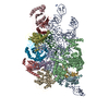
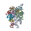
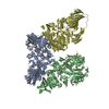
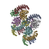
| Title | Cryo-EM structure of yeast Ribonuclease P | |||||||||
 Map data Map data | ||||||||||
 Sample Sample |
| |||||||||
 Keywords Keywords | Ribonuclease P / RNA-protein complex / HYDROLASE-RNA complex | |||||||||
| Function / homology |  Function and homology information Function and homology informationribonuclease MRP activity / nuclear-transcribed mRNA catabolic process, RNase MRP-dependent / intronic box C/D snoRNA processing / nucleolar ribonuclease P complex / ribonuclease MRP complex / ribonuclease P RNA binding / ribonuclease P complex / ribonuclease P / rRNA primary transcript binding / ribonuclease P activity ...ribonuclease MRP activity / nuclear-transcribed mRNA catabolic process, RNase MRP-dependent / intronic box C/D snoRNA processing / nucleolar ribonuclease P complex / ribonuclease MRP complex / ribonuclease P RNA binding / ribonuclease P complex / ribonuclease P / rRNA primary transcript binding / ribonuclease P activity / tRNA 5'-leader removal / telomerase holoenzyme complex / tRNA processing / maturation of 5.8S rRNA / rRNA processing / RNA binding / metal ion binding / nucleus / cytoplasm / cytosol Similarity search - Function | |||||||||
| Biological species |  | |||||||||
| Method | single particle reconstruction / cryo EM / Resolution: 3.48 Å | |||||||||
 Authors Authors | Lan P / Tan M | |||||||||
 Citation Citation |  Journal: Science / Year: 2018 Journal: Science / Year: 2018Title: Structural insight into precursor tRNA processing by yeast ribonuclease P. Authors: Pengfei Lan / Ming Tan / Yuebin Zhang / Shuangshuang Niu / Juan Chen / Shaohua Shi / Shuwan Qiu / Xuejuan Wang / Xiangda Peng / Gang Cai / Hong Cheng / Jian Wu / Guohui Li / Ming Lei /  Abstract: Ribonuclease P (RNase P) is a universal ribozyme responsible for processing the 5'-leader of pre-transfer RNA (pre-tRNA). Here, we report the 3.5-angstrom cryo-electron microscopy structures of ...Ribonuclease P (RNase P) is a universal ribozyme responsible for processing the 5'-leader of pre-transfer RNA (pre-tRNA). Here, we report the 3.5-angstrom cryo-electron microscopy structures of RNase P alone and in complex with pre-tRNA The protein components form a hook-shaped architecture that wraps around the RNA and stabilizes RNase P into a "measuring device" with two fixed anchors that recognize the L-shaped pre-tRNA. A universally conserved uridine nucleobase and phosphate backbone in the catalytic center together with the scissile phosphate and the O3' leaving group of pre-tRNA jointly coordinate two catalytic magnesium ions. Binding of pre-tRNA induces a conformational change in the catalytic center that is required for catalysis. Moreover, simulation analysis suggests a two-metal-ion S2 reaction pathway of pre-tRNA cleavage. These results not only reveal the architecture of yeast RNase P but also provide a molecular basis of how the 5'-leader of pre-tRNA is processed by eukaryotic RNase P. | |||||||||
| History |
|
- Structure visualization
| Movie |
 Movie viewer Movie viewer |
|---|---|
| Structure viewer | EM map:  SurfView SurfView Molmil Molmil Jmol/JSmol Jmol/JSmol |
| Supplemental images |
- Downloads & links
-EMDB archive
| Map data |  emd_9616.map.gz emd_9616.map.gz | 59.9 MB |  EMDB map data format EMDB map data format | |
|---|---|---|---|---|
| Header (meta data) |  emd-9616-v30.xml emd-9616-v30.xml emd-9616.xml emd-9616.xml | 20.5 KB 20.5 KB | Display Display |  EMDB header EMDB header |
| Images |  emd_9616.png emd_9616.png | 76.8 KB | ||
| Filedesc metadata |  emd-9616.cif.gz emd-9616.cif.gz | 7 KB | ||
| Archive directory |  http://ftp.pdbj.org/pub/emdb/structures/EMD-9616 http://ftp.pdbj.org/pub/emdb/structures/EMD-9616 ftp://ftp.pdbj.org/pub/emdb/structures/EMD-9616 ftp://ftp.pdbj.org/pub/emdb/structures/EMD-9616 | HTTPS FTP |
-Related structure data
| Related structure data |  6agbMC  9622C  6ah3C M: atomic model generated by this map C: citing same article ( |
|---|---|
| Similar structure data |
- Links
Links
| EMDB pages |  EMDB (EBI/PDBe) / EMDB (EBI/PDBe) /  EMDataResource EMDataResource |
|---|---|
| Related items in Molecule of the Month |
- Map
Map
| File |  Download / File: emd_9616.map.gz / Format: CCP4 / Size: 64 MB / Type: IMAGE STORED AS FLOATING POINT NUMBER (4 BYTES) Download / File: emd_9616.map.gz / Format: CCP4 / Size: 64 MB / Type: IMAGE STORED AS FLOATING POINT NUMBER (4 BYTES) | ||||||||||||||||||||||||||||||||||||||||||||||||||||||||||||
|---|---|---|---|---|---|---|---|---|---|---|---|---|---|---|---|---|---|---|---|---|---|---|---|---|---|---|---|---|---|---|---|---|---|---|---|---|---|---|---|---|---|---|---|---|---|---|---|---|---|---|---|---|---|---|---|---|---|---|---|---|---|
| Projections & slices | Image control
Images are generated by Spider. | ||||||||||||||||||||||||||||||||||||||||||||||||||||||||||||
| Voxel size | X=Y=Z: 1.32 Å | ||||||||||||||||||||||||||||||||||||||||||||||||||||||||||||
| Density |
| ||||||||||||||||||||||||||||||||||||||||||||||||||||||||||||
| Symmetry | Space group: 1 | ||||||||||||||||||||||||||||||||||||||||||||||||||||||||||||
| Details | EMDB XML:
CCP4 map header:
| ||||||||||||||||||||||||||||||||||||||||||||||||||||||||||||
-Supplemental data
- Sample components
+Entire : RNase P
+Supramolecule #1: RNase P
+Macromolecule #1: Ribonuclease P RNA
+Macromolecule #2: Ribonucleases P/MRP protein subunit POP1
+Macromolecule #3: Ribonucleases P/MRP protein subunit POP3
+Macromolecule #4: RNases MRP/P 32.9 kDa subunit
+Macromolecule #5: Ribonuclease P/MRP protein subunit POP5
+Macromolecule #6: Ribonucleases P/MRP protein subunit POP6
+Macromolecule #7: Ribonucleases P/MRP protein subunit POP7
+Macromolecule #8: Ribonucleases P/MRP protein subunit POP8
+Macromolecule #9: Ribonuclease P/MRP protein subunit RPP1
+Macromolecule #10: Ribonuclease P protein subunit RPR2
+Macromolecule #11: ZINC ION
-Experimental details
-Structure determination
| Method | cryo EM |
|---|---|
 Processing Processing | single particle reconstruction |
| Aggregation state | particle |
- Sample preparation
| Buffer | pH: 7.5 |
|---|---|
| Vitrification | Cryogen name: ETHANE |
- Electron microscopy
| Microscope | FEI TITAN KRIOS |
|---|---|
| Image recording | Film or detector model: GATAN K2 SUMMIT (4k x 4k) / Average electron dose: 50.0 e/Å2 |
| Electron beam | Acceleration voltage: 300 kV / Electron source:  FIELD EMISSION GUN FIELD EMISSION GUN |
| Electron optics | Illumination mode: FLOOD BEAM / Imaging mode: DIFFRACTION |
| Experimental equipment |  Model: Titan Krios / Image courtesy: FEI Company |
- Image processing
| Startup model | Type of model: NONE |
|---|---|
| Final reconstruction | Resolution.type: BY AUTHOR / Resolution: 3.48 Å / Resolution method: FSC 0.143 CUT-OFF / Number images used: 164765 |
| Initial angle assignment | Type: MAXIMUM LIKELIHOOD |
| Final angle assignment | Type: MAXIMUM LIKELIHOOD |
 Movie
Movie Controller




 Z (Sec.)
 Y (Row.)
 X (Col.)


