 Movie
Movie Controller

[English] 日本語
 Yorodumi
- PDB-6yyk: Crystal Structure of 1,5-dimethylindoline-2,3-dione covalently bo... -
+ Open data

- Basic information
| Entry | Database: PDB / ID: 6yyk | ||||||
|---|---|---|---|---|---|---|---|
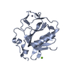
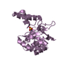
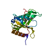
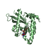
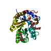
| Title | Crystal Structure of 1,5-dimethylindoline-2,3-dione covalently bound to the PH domain of Bruton's tyrosine kinase mutant R28C | ||||||
 Components Components | Tyrosine-protein kinase BTK | ||||||
 Keywords Keywords | TRANSFERASE / BTK / Covalent fragments / surface entrophy reduction / crystal engineering | ||||||
| Function / homology |  Function and homology information Function and homology informationregulation of B cell cytokine production / regulation of B cell apoptotic process / monocyte proliferation / positive regulation of interleukin-17A production / proteoglycan catabolic process / eosinophil homeostasis / positive regulation of type III hypersensitivity / B cell affinity maturation / positive regulation of synoviocyte proliferation / histamine secretion by mast cell ...regulation of B cell cytokine production / regulation of B cell apoptotic process / monocyte proliferation / positive regulation of interleukin-17A production / proteoglycan catabolic process / eosinophil homeostasis / positive regulation of type III hypersensitivity / B cell affinity maturation / positive regulation of synoviocyte proliferation / histamine secretion by mast cell / positive regulation of cGAS/STING signaling pathway / neutrophil homeostasis / positive regulation of type I hypersensitivity / cellular response to molecule of fungal origin / MyD88 deficiency (TLR2/4) / cellular response to interleukin-7 / IRAK4 deficiency (TLR2/4) / MyD88:MAL(TIRAP) cascade initiated on plasma membrane / MyD88-dependent toll-like receptor signaling pathway / positive regulation of B cell differentiation / phospholipase activator activity / negative regulation of interleukin-10 production / negative regulation of B cell proliferation / positive regulation of immunoglobulin production / Fc-epsilon receptor signaling pathway / mesoderm development / phosphatidylinositol-3,4,5-trisphosphate binding / positive regulation of NLRP3 inflammasome complex assembly / B cell activation / RHO GTPases Activate WASPs and WAVEs / phospholipase binding / positive regulation of B cell proliferation / peptidyl-tyrosine phosphorylation / cell maturation / FCERI mediated Ca+2 mobilization / positive regulation of phagocytosis / Antigen activates B Cell Receptor (BCR) leading to generation of second messengers / : / B cell receptor signaling pathway / non-specific protein-tyrosine kinase / FCGR3A-mediated phagocytosis / cellular response to reactive oxygen species / non-membrane spanning protein tyrosine kinase activity / apoptotic signaling pathway / calcium-mediated signaling / Regulation of actin dynamics for phagocytic cup formation / positive regulation of interleukin-6 production / positive regulation of tumor necrosis factor production / G beta:gamma signalling through BTK / DAP12 signaling / T cell receptor signaling pathway / G alpha (12/13) signalling events / ER-Phagosome pathway / protein tyrosine kinase activity / cytoplasmic vesicle / response to lipopolysaccharide / Potential therapeutics for SARS / G alpha (q) signalling events / adaptive immune response / positive regulation of canonical NF-kappaB signal transduction / intracellular signal transduction / membrane raft / innate immune response / perinuclear region of cytoplasm / zinc ion binding / ATP binding / identical protein binding / nucleus / plasma membrane / cytoplasm / cytosol Similarity search - Function | ||||||
| Biological species |  Homo sapiens (human) Homo sapiens (human) | ||||||
| Method |  X-RAY DIFFRACTION / SYNCHROTRON / MOLECULAR REPLACEMENT / molecular replacement / Resolution: 2.04 Å X-RAY DIFFRACTION / SYNCHROTRON / MOLECULAR REPLACEMENT / molecular replacement / Resolution: 2.04 Å | ||||||
 Authors Authors | Brear, P. / Wagstaff, J. / Hyvonen, M. | ||||||
| Funding support |  United Kingdom, 1items United Kingdom, 1items
| ||||||
 Citation Citation | Journal: To Be Published Title: Optimising crystallographic systems for structure-guided drug discovery Authors: Brear, P. / Fischer, G. / May, M. / Pantelejevs, T. / Mathieu, R. / Rossmann, M. / Wagstaff, J. / Blaszczyk, B. / Hyvonen, M. | ||||||
| History |
|
- Structure visualization
| Structure viewer | Molecule: MolmilJmol/JSmol |
|---|
- Downloads & links
-Download
| PDBx/mmCIF format | 6yyk.cif.gz | 81.9 KB | Display | PDBx/mmCIF format |
|---|---|---|---|---|
| PDB format | pdb6yyk.ent.gz | 60 KB | Display | PDB format |
| PDBx/mmJSON format | 6yyk.json.gz | Tree view | PDBx/mmJSON format | |
| Others |  Other downloads Other downloads |
-Validation report
| Arichive directory | https://data.pdbj.org/pub/pdb/validation_reports/yy/6yykftp://data.pdbj.org/pub/pdb/validation_reports/yy/6yyk | HTTPS FTP |
|---|
-Related structure data
| Related structure data |  6xufC  6xujC  6yyfC  6yygC  1btkS S: Starting model for refinement C: citing same article ( |
|---|---|
| Similar structure data |


-Links
 PDBj
PDBj


- Assembly
| Deposited unit | 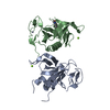
| ||||||||
|---|---|---|---|---|---|---|---|---|---|
| 1 | 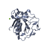
| ||||||||
| 2 | 
| ||||||||
| Unit cell |
|
-Components
| #1: Protein | Mass: 19928.963 Da / Num. of mol.: 2 Source method: isolated from a genetically manipulated source Source: (gene. exp.) Homo sapiens (human) / Gene: BTK, AGMX1, ATK, BPK / Plasmid: pBAT4 / Production host:  References: UniProt: Q06187, non-specific protein-tyrosine kinase #2: Chemical |   Mass: 65.409 Da / Num. of mol.: 2 / Source method: obtained synthetically / Formula: Zn Mass: 65.409 Da / Num. of mol.: 2 / Source method: obtained synthetically / Formula: Zn#3: Chemical |   Mass: 24.305 Da / Num. of mol.: 2 / Source method: obtained synthetically / Formula: Mg Mass: 24.305 Da / Num. of mol.: 2 / Source method: obtained synthetically / Formula: Mg#4: Chemical | ChemComp-IS7 / | 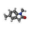  Mass: 161.200 Da / Num. of mol.: 1 / Source method: obtained synthetically / Formula: C10H11NO / Feature type: SUBJECT OF INVESTIGATION Mass: 161.200 Da / Num. of mol.: 1 / Source method: obtained synthetically / Formula: C10H11NO / Feature type: SUBJECT OF INVESTIGATION#5: Water | ChemComp-HOH / |  Mass: 18.015 Da / Num. of mol.: 30 / Source method: isolated from a natural source / Formula: H2O Mass: 18.015 Da / Num. of mol.: 30 / Source method: isolated from a natural source / Formula: H2OHas ligand of interest | Y | Has protein modification | Y | |
|---|
-Experimental details
-Experiment
| Experiment | Method: X-RAY DIFFRACTION / Number of used crystals: 1 |
|---|
- Sample preparation
| Crystal | Density Matthews: 2.1 Å3/Da / Density % sol: 41.55 % / Mosaicity: 0 ° |
|---|---|
| Crystal grow | Temperature: 298 K / Method: vapor diffusion, sitting drop / pH: 8.5 Details: 0.1 M TRIS 8.5 pH, 32.5% w/v PEG 3350, 200mM MgCl2 500 mM NaCl |
-Data collection
| Diffraction | Mean temperature: 100 K / Serial crystal experiment: N | |||||||||||||||||||||||||||
|---|---|---|---|---|---|---|---|---|---|---|---|---|---|---|---|---|---|---|---|---|---|---|---|---|---|---|---|---|
| Diffraction source | Source: SYNCHROTRON / Site: Diamond / Beamline: I04 / Wavelength: 0.9795 Å | |||||||||||||||||||||||||||
| Detector | Type: DECTRIS PILATUS 6M / Detector: PIXEL / Date: Apr 3, 2017 | |||||||||||||||||||||||||||
| Radiation | Protocol: SINGLE WAVELENGTH / Monochromatic (M) / Laue (L): M / Scattering type: x-ray | |||||||||||||||||||||||||||
| Radiation wavelength | Wavelength: 0.9795 Å / Relative weight: 1 | |||||||||||||||||||||||||||
| Reflection | Resolution: 2.04→60.41 Å / Num. obs: 21231 / % possible obs: 99.8 % / Redundancy: 3.2 % / CC1/2: 0.996 / Rmerge(I) obs: 0.074 / Rpim(I) all: 0.049 / Rrim(I) all: 0.09 / Net I/σ(I): 5.3 / Num. measured all: 67401 / Scaling rejects: 11 | |||||||||||||||||||||||||||
| Reflection shell | Diffraction-ID: 1 / Redundancy: 3 %
|
-Phasing
| Phasing | Method: molecular replacement |
|---|
- Processing
| Software |
| ||||||||||||||||||||||||||||||||||||||||||||||||||||||||||||
|---|---|---|---|---|---|---|---|---|---|---|---|---|---|---|---|---|---|---|---|---|---|---|---|---|---|---|---|---|---|---|---|---|---|---|---|---|---|---|---|---|---|---|---|---|---|---|---|---|---|---|---|---|---|---|---|---|---|---|---|---|---|
| Refinement | Method to determine structure: MOLECULAR REPLACEMENT Starting model: 1BTK Resolution: 2.04→57.74 Å / Cor.coef. Fo:Fc: 0.961 / Cor.coef. Fo:Fc free: 0.931 / SU B: 15.021 / SU ML: 0.333 / Cross valid method: THROUGHOUT / σ(F): 0 / ESU R: 0.259 / ESU R Free: 0.228 / Stereochemistry target values: MAXIMUM LIKELIHOOD Details: HYDROGENS HAVE BEEN ADDED IN THE RIDING POSITIONS U VALUES : REFINED INDIVIDUALLY
| ||||||||||||||||||||||||||||||||||||||||||||||||||||||||||||
| Solvent computation | Ion probe radii: 0.8 Å / Shrinkage radii: 0.8 Å / VDW probe radii: 1.2 Å / Solvent model: MASK | ||||||||||||||||||||||||||||||||||||||||||||||||||||||||||||
| Displacement parameters | Biso max: 203.38 Å2 / Biso mean: 67.47 Å2 / Biso min: 36.46 Å2
| ||||||||||||||||||||||||||||||||||||||||||||||||||||||||||||
| Refinement step | Cycle: final / Resolution: 2.04→57.74 Å
| ||||||||||||||||||||||||||||||||||||||||||||||||||||||||||||
| Refine LS restraints |
| ||||||||||||||||||||||||||||||||||||||||||||||||||||||||||||
| LS refinement shell | Resolution: 2.04→2.089 Å / Rfactor Rfree error: 0
|
