- PDB-6ysh: Lamin A 1-70 coil1A dimer stabilized by C-terminal capping -
+
Open data
ID or keywords:
Loading...
-
Basic information
Entry
Database: PDB / ID: 6ysh
Title
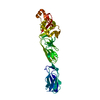
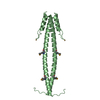
Lamin A 1-70 coil1A dimer stabilized by C-terminal capping
Components
(Prelamin-A/C,Microtubule-associated protein RP/EB family member 1) x 2
Keywords
NUCLEAR PROTEIN / intermediate filaments lamin coiled-coil
Function / homology
Function and homology information
structural constituent of nuclear lamina / negative regulation of mesenchymal cell proliferation / protein localization to astral microtubule / protein localization to mitotic spindle / cortical microtubule cytoskeleton / mitotic spindle astral microtubule end / protein localization to nuclear envelope / establishment or maintenance of microtubule cytoskeleton polarity / Breakdown of the nuclear lamina / ventricular cardiac muscle cell development ...structural constituent of nuclear lamina / negative regulation of mesenchymal cell proliferation / protein localization to astral microtubule / protein localization to mitotic spindle / cortical microtubule cytoskeleton / mitotic spindle astral microtubule end / protein localization to nuclear envelope / establishment or maintenance of microtubule cytoskeleton polarity / Breakdown of the nuclear lamina / ventricular cardiac muscle cell development / Depolymerization of the Nuclear Lamina / Nuclear Envelope Breakdown / nuclear pore localization / DNA double-strand break attachment to nuclear envelope / protein localization to microtubule / lamin filament / nuclear envelope organization / cell projection membrane / microtubule plus-end / mitotic spindle microtubule / XBP1(S) activates chaperone genes / nuclear lamina / attachment of mitotic spindle microtubules to kinetochore / microtubule plus-end binding / microtubule bundle formation / non-motile cilium assembly / Initiation of Nuclear Envelope (NE) Reformation / regulation of protein localization to nucleus / protein localization to centrosome / regulation of telomere maintenance / intermediate filament / negative regulation of cardiac muscle hypertrophy in response to stress / muscle organ development / nuclear migration / Deregulated CDK5 triggers multiple neurodegenerative pathways in Alzheimer's disease models / mitotic spindle pole / negative regulation of release of cytochrome c from mitochondria / spindle midzone / negative regulation of microtubule polymerization / establishment of mitotic spindle orientation / microtubule polymerization / microtubule organizing center / regulation of microtubule polymerization or depolymerization / protein localization to nucleus / positive regulation of microtubule polymerization / cytoplasmic microtubule / spindle assembly / Amplification of signal from unattached kinetochores via a MAD2 inhibitory signal / Loss of Nlp from mitotic centrosomes / Loss of proteins required for interphase microtubule organization from the centrosome / Recruitment of mitotic centrosome proteins and complexes / Recruitment of NuMA to mitotic centrosomes / Anchoring of the basal body to the plasma membrane / Mitotic Prometaphase / EML4 and NUDC in mitotic spindle formation / regulation of cell migration / AURKA Activation by TPX2 / Meiotic synapsis / Resolution of Sister Chromatid Cohesion / negative regulation of extrinsic apoptotic signaling pathway / protein serine/threonine kinase binding / regulation of protein stability / RHO GTPases Activate Formins / structural constituent of cytoskeleton / double-strand break repair via nonhomologous end joining / nuclear matrix / protein import into nucleus / cellular senescence / The role of GTSE1 in G2/M progression after G2 checkpoint / Signaling by BRAF and RAF1 fusions / Separation of Sister Chromatids / intracellular protein localization / Regulation of PLK1 Activity at G2/M Transition / nuclear envelope / cell migration / heterochromatin formation / site of double-strand break / nuclear membrane / cellular response to hypoxia / microtubule / nuclear speck / ciliary basal body / cadherin binding / negative regulation of cell population proliferation / cell division / focal adhesion / positive regulation of gene expression / centrosome / perinuclear region of cytoplasm / structural molecule activity / Golgi apparatus / RNA binding / nucleoplasm / identical protein binding / nucleus / cytosol Similarity search - Function
Single alpha-helices involved in coiled-coils or other helix-helix interfaces - #1430 / Lamin tail domain superfamily / Lamin tail domain / Lamin Tail Domain / Lamin-tail (LTD) domain profile. / EB1, C-terminal / Microtubule-associated protein RP/EB / EB1, C-terminal domain superfamily / EB1-C terminal (EB1-C) domain profile. / EB1-like C-terminal motif ...Single alpha-helices involved in coiled-coils or other helix-helix interfaces - #1430 / Lamin tail domain superfamily / Lamin tail domain / Lamin Tail Domain / Lamin-tail (LTD) domain profile. / EB1, C-terminal / Microtubule-associated protein RP/EB / EB1, C-terminal domain superfamily / EB1-C terminal (EB1-C) domain profile. / EB1-like C-terminal motif / Intermediate filament protein, conserved site / Intermediate filament protein / Intermediate filament (IF) rod domain signature. / Intermediate filament, rod domain / Intermediate filament (IF) rod domain profile. / Intermediate filament protein / Calponin homology (CH) domain / Calponin homology domain / CH domain superfamily / Calponin homology (CH) domain profile. / Single alpha-helices involved in coiled-coils or other helix-helix interfaces / Up-down Bundle / Mainly Alpha Similarity search - Domain/homology
Resolution: 2.83→83.58 Å / Cor.coef. Fo:Fc: 0.951 / Cor.coef. Fo:Fc free: 0.93 / SU B: 48.452 / SU ML: 0.396 / Cross valid method: THROUGHOUT / ESU R: 0.422 / ESU R Free: 0.315 / Stereochemistry target values: MAXIMUM LIKELIHOOD / Details: HYDROGENS HAVE BEEN ADDED IN THE RIDING POSITIONS
Rfactor
Num. reflection
% reflection
Selection details
Rfree
0.28131
496
5.1 %
RANDOM
Rwork
0.23747
-
-
-
obs
0.23972
9158
99.81 %
-
Solvent computation
Ion probe radii: 0.8 Å / Shrinkage radii: 0.8 Å / VDW probe radii: 1.2 Å / Solvent model: MASK
Displacement parameters
Biso mean: 110.135 Å2
Baniso -1
Baniso -2
Baniso -3
1-
-8.1 Å2
0 Å2
-0 Å2
2-
-
-8.1 Å2
-0 Å2
3-
-
-
16.21 Å2
Refinement step
Cycle: 1 / Resolution: 2.83→83.58 Å
Protein
Nucleic acid
Ligand
Solvent
Total
Num. atoms
1360
0
0
22
1382
Refine LS restraints
Refine-ID
Type
Dev ideal
Dev ideal target
Number
X-RAY DIFFRACTION
r_bond_refined_d
0.018
0.013
1371
X-RAY DIFFRACTION
r_bond_other_d
0.003
0.017
1294
X-RAY DIFFRACTION
r_angle_refined_deg
1.909
1.65
1848
X-RAY DIFFRACTION
r_angle_other_deg
1.505
1.583
2988
X-RAY DIFFRACTION
r_dihedral_angle_1_deg
3.687
5
162
X-RAY DIFFRACTION
r_dihedral_angle_2_deg
35.526
22.277
101
X-RAY DIFFRACTION
r_dihedral_angle_3_deg
18.705
15
265
X-RAY DIFFRACTION
r_dihedral_angle_4_deg
25.033
15
17
X-RAY DIFFRACTION
r_chiral_restr
0.106
0.2
180
X-RAY DIFFRACTION
r_gen_planes_refined
0.007
0.02
1542
X-RAY DIFFRACTION
r_gen_planes_other
0.002
0.02
289
X-RAY DIFFRACTION
r_nbd_refined
X-RAY DIFFRACTION
r_nbd_other
X-RAY DIFFRACTION
r_nbtor_refined
X-RAY DIFFRACTION
r_nbtor_other
X-RAY DIFFRACTION
r_xyhbond_nbd_refined
X-RAY DIFFRACTION
r_xyhbond_nbd_other
X-RAY DIFFRACTION
r_metal_ion_refined
X-RAY DIFFRACTION
r_metal_ion_other
X-RAY DIFFRACTION
r_symmetry_vdw_refined
X-RAY DIFFRACTION
r_symmetry_vdw_other
X-RAY DIFFRACTION
r_symmetry_hbond_refined
X-RAY DIFFRACTION
r_symmetry_hbond_other
X-RAY DIFFRACTION
r_symmetry_metal_ion_refined
X-RAY DIFFRACTION
r_symmetry_metal_ion_other
X-RAY DIFFRACTION
r_mcbond_it
8.644
9.074
654
X-RAY DIFFRACTION
r_mcbond_other
8.645
9.068
653
X-RAY DIFFRACTION
r_mcangle_it
12.417
13.607
814
X-RAY DIFFRACTION
r_mcangle_other
12.41
13.614
815
X-RAY DIFFRACTION
r_scbond_it
10.662
10.572
717
X-RAY DIFFRACTION
r_scbond_other
10.672
10.562
715
X-RAY DIFFRACTION
r_scangle_it
X-RAY DIFFRACTION
r_scangle_other
16.827
15.37
1034
X-RAY DIFFRACTION
r_rigid_bond_restr
X-RAY DIFFRACTION
r_sphericity_free
X-RAY DIFFRACTION
r_sphericity_bonded
LS refinement shell
Resolution: 2.83→2.904 Å / Total num. of bins used: 20
Rfactor
Num. reflection
% reflection
Rfree
0.419
39
-
Rwork
0.422
639
-
obs
-
-
99.12 %
Refinement TLS params.
Method: refined / Refine-ID: X-RAY DIFFRACTION
ID
L11 (°2)
L12 (°2)
L13 (°2)
L22 (°2)
L23 (°2)
L33 (°2)
S11 (Å °)
S12 (Å °)
S13 (Å °)
S21 (Å °)
S22 (Å °)
S23 (Å °)
S31 (Å °)
S32 (Å °)
S33 (Å °)
T11 (Å2)
T12 (Å2)
T13 (Å2)
T22 (Å2)
T23 (Å2)
T33 (Å2)
Origin x (Å)
Origin y (Å)
Origin z (Å)
1
4.5615
-1.4879
0.1825
1.108
-0.0813
0.0777
-0.0063
-0.1094
0.5139
-0.0872
0.0594
-0.2149
-0.0966
0.0043
-0.053
0.1568
-0.0057
0.112
0.0857
0.0185
0.1541
21.5089
9.51
-7.187
2
0.3653
0.3827
-0.113
0.4495
-0.1152
0.0391
-0.18
0.1024
-0.1484
-0.1327
0.1433
-0.0791
0.0291
-0.0389
0.0366
0.1584
0.0451
0.0182
0.1563
0.0143
0.2519
20.067
11.3854
-5.3879
Refinement TLS group
ID
Refine-ID
Refine TLS-ID
Auth asym-ID
Auth seq-ID
1
X-RAY DIFFRACTION
1
A
25 - 107
2
X-RAY DIFFRACTION
2
B
26 - 106
+
About Yorodumi
-
News
-
Feb 9, 2022. New format data for meta-information of EMDB entries
New format data for meta-information of EMDB entries
Version 3 of the EMDB header file is now the official format.
The previous official version 1.9 will be removed from the archive.
In the structure databanks used in Yorodumi, some data are registered as the other names, "COVID-19 virus" and "2019-nCoV". Here are the details of the virus and the list of structure data.
Jan 31, 2019. EMDB accession codes are about to change! (news from PDBe EMDB page)
EMDB accession codes are about to change! (news from PDBe EMDB page)
The allocation of 4 digits for EMDB accession codes will soon come to an end. Whilst these codes will remain in use, new EMDB accession codes will include an additional digit and will expand incrementally as the available range of codes is exhausted. The current 4-digit format prefixed with “EMD-” (i.e. EMD-XXXX) will advance to a 5-digit format (i.e. EMD-XXXXX), and so on. It is currently estimated that the 4-digit codes will be depleted around Spring 2019, at which point the 5-digit format will come into force.
The EM Navigator/Yorodumi systems omit the EMD- prefix.
Related info.:Q: What is EMD? / ID/Accession-code notation in Yorodumi/EM Navigator
Yorodumi is a browser for structure data from EMDB, PDB, SASBDB, etc.
This page is also the successor to EM Navigator detail page, and also detail information page/front-end page for Omokage search.
The word "yorodu" (or yorozu) is an old Japanese word meaning "ten thousand". "mi" (miru) is to see.
Related info.:EMDB / PDB / SASBDB / Comparison of 3 databanks / Yorodumi Search / Aug 31, 2016. New EM Navigator & Yorodumi / Yorodumi Papers / Jmol/JSmol / Function and homology information / Changes in new EM Navigator and Yorodumi
 Movie
Movie Controller


 Open data
 Basic information
 Components
 Keywords
 Function and homology information
 Homo sapiens (human)
 X-RAY DIFFRACTION /
  Authors
 Belgium, 1items
  Citation
 Structure visualization
 Downloads & links
 Other downloads


 PDBj
PDBj


 Assembly

 Mass: 18.015 Da / Num. of mol.: 22 / Source method: isolated from a natural source / Formula: H2O
 Sample preparation
 / Beamline: P14 (MX2) / Wavelength: 0.97631 Å
 Processing