 Movie
Movie Controller


+ Open data

- Basic information
| Entry | Database: PDB / ID: 6yjd | ||||||
|---|---|---|---|---|---|---|---|
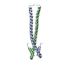
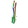
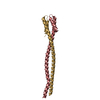
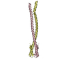
| Title | Lamin A coil2 dimer stabilized by N-terminal capping | ||||||
 Components Components | Capsid assembly scaffolding protein,Prelamin-A/C | ||||||
 Keywords Keywords | NUCLEAR PROTEIN / intermediate filaments / lamin / coiled-coil | ||||||
| Function / homology |  Function and homology information Function and homology informationviral scaffold / structural constituent of nuclear lamina / protein localization to nuclear envelope / establishment or maintenance of microtubule cytoskeleton polarity / Breakdown of the nuclear lamina / Depolymerization of the Nuclear Lamina / Nuclear Envelope Breakdown / nuclear pore localization / DNA double-strand break attachment to nuclear envelope / lamin filament ...viral scaffold / structural constituent of nuclear lamina / protein localization to nuclear envelope / establishment or maintenance of microtubule cytoskeleton polarity / Breakdown of the nuclear lamina / Depolymerization of the Nuclear Lamina / Nuclear Envelope Breakdown / nuclear pore localization / DNA double-strand break attachment to nuclear envelope / lamin filament / nuclear envelope organization / XBP1(S) activates chaperone genes / nuclear lamina / Initiation of Nuclear Envelope (NE) Reformation / regulation of telomere maintenance / intermediate filament / negative regulation of cardiac muscle hypertrophy in response to stress / muscle organ development / nuclear migration / virion assembly / Deregulated CDK5 triggers multiple neurodegenerative pathways in Alzheimer's disease models / protein localization to nucleus / regulation of cell migration / Meiotic synapsis / structural constituent of cytoskeleton / double-strand break repair via nonhomologous end joining / cellular senescence / nuclear matrix / Signaling by BRAF and RAF1 fusions / intracellular protein localization / nuclear envelope / heterochromatin formation / site of double-strand break / nuclear membrane / cellular response to hypoxia / nuclear speck / negative regulation of cell population proliferation / perinuclear region of cytoplasm / structural molecule activity / DNA binding / nucleoplasm / identical protein binding / nucleus / cytosol Similarity search - Function | ||||||
| Biological species |   Bacillus phage phi29 (virus) Bacillus phage phi29 (virus) Homo sapiens (human) Homo sapiens (human) | ||||||
| Method |  X-RAY DIFFRACTION / SYNCHROTRON / SAD / Resolution: 2.9 Å X-RAY DIFFRACTION / SYNCHROTRON / SAD / Resolution: 2.9 Å | ||||||
 Authors Authors | Lilina, A.V. / Stalmans, G. / Strelkov, S.V. | ||||||
| Funding support |  Belgium, 1items Belgium, 1items
| ||||||
 Citation Citation | Journal: Cells / Year: 2020 Title: Addressing the Molecular Mechanism of Longitudinal Lamin Assembly Using Chimeric Fusions. Authors: Stalmans, G. / Lilina, A.V. / Vermeire, P.J. / Fiala, J. / Novak, P. / Strelkov, S.V. | ||||||
| History |
|
- Structure visualization
| Structure viewer | Molecule: MolmilJmol/JSmol |
|---|
- Downloads & links
-Download
| PDBx/mmCIF format | 6yjd.cif.gz | 37.6 KB | Display | PDBx/mmCIF format |
|---|---|---|---|---|
| PDB format | pdb6yjd.ent.gz | 22.8 KB | Display | PDB format |
| PDBx/mmJSON format | 6yjd.json.gz | Tree view | PDBx/mmJSON format | |
| Others |  Other downloads Other downloads |
-Validation report
| Arichive directory | https://data.pdbj.org/pub/pdb/validation_reports/yj/6yjdftp://data.pdbj.org/pub/pdb/validation_reports/yj/6yjd | HTTPS FTP |
|---|
-Related structure data


-Links
 PDBj
PDBj


- Assembly
| Deposited unit | 
| ||||||||||
|---|---|---|---|---|---|---|---|---|---|---|---|
| 1 | 
| ||||||||||
| Unit cell |
|
-Components
| #1: Protein | Mass: 14894.788 Da / Num. of mol.: 1 / Mutation: F40C Source method: isolated from a genetically manipulated source Source: (gene. exp.) Bacillus phage phi29 (virus), (gene. exp.) Homo sapiens (human)Gene: LMNA, LMN1 / Production host:  | ||||||||||
|---|---|---|---|---|---|---|---|---|---|---|---|
| #2: Chemical |   Mass: 58.693 Da / Num. of mol.: 2 / Source method: obtained synthetically / Formula: Ni Mass: 58.693 Da / Num. of mol.: 2 / Source method: obtained synthetically / Formula: Ni#3: Chemical | ChemComp-CL / |   Mass: 35.453 Da / Num. of mol.: 1 / Source method: obtained synthetically / Formula: Cl Mass: 35.453 Da / Num. of mol.: 1 / Source method: obtained synthetically / Formula: Cl#4: Chemical |   Mass: 122.143 Da / Num. of mol.: 2 / Source method: obtained synthetically / Formula: C4H12NO3 / Comment: pH buffer*YM Mass: 122.143 Da / Num. of mol.: 2 / Source method: obtained synthetically / Formula: C4H12NO3 / Comment: pH buffer*YM#5: Water | ChemComp-HOH / |  Mass: 18.015 Da / Num. of mol.: 2 / Source method: isolated from a natural source / Formula: H2O Mass: 18.015 Da / Num. of mol.: 2 / Source method: isolated from a natural source / Formula: H2OHas ligand of interest | N | Has protein modification | Y | |
-Experimental details
-Experiment
| Experiment | Method: X-RAY DIFFRACTION / Number of used crystals: 1 |
|---|
- Sample preparation
| Crystal | Density Matthews: 6.21 Å3/Da / Density % sol: 80.18 % |
|---|---|
| Crystal grow | Temperature: 277 K / Method: vapor diffusion, hanging drop / pH: 8.2 / Details: 35% (v/v) methanol, 0.2 M MgCl2 and 0.1 M HEPES |
-Data collection
| Diffraction | Mean temperature: 100 K / Serial crystal experiment: N |
|---|---|
| Diffraction source | Source: SYNCHROTRON / Site: SOLEIL  / Beamline: PROXIMA 1 / Wavelength: 0.978565 Å / Beamline: PROXIMA 1 / Wavelength: 0.978565 Å |
| Detector | Type: DECTRIS EIGER X 16M / Detector: PIXEL / Date: Feb 15, 2019 |
| Radiation | Protocol: MAD / Monochromatic (M) / Laue (L): M / Scattering type: x-ray |
| Radiation wavelength | Wavelength: 0.978565 Å / Relative weight: 1 |
| Reflection | Resolution: 2.9→44.62 Å / Num. obs: 8792 / % possible obs: 98.28 % / Redundancy: 19.2 % / Biso Wilson estimate: 110.62 Å2 / CC1/2: 0.999 / Net I/σ(I): 20.27 |
| Reflection shell | Resolution: 2.9→3 Å / Mean I/σ(I) obs: 0.99 / Num. unique obs: 847 / CC1/2: 0.934 |
- Processing
| Software |
| ||||||||||||||||||||||||||||||||||||||||||||||||||||||||||||||||||||||||||||||||||||||||||||||||||||||||||||||||||||||||||||||||||||||||||||||||||||||||||||||||
|---|---|---|---|---|---|---|---|---|---|---|---|---|---|---|---|---|---|---|---|---|---|---|---|---|---|---|---|---|---|---|---|---|---|---|---|---|---|---|---|---|---|---|---|---|---|---|---|---|---|---|---|---|---|---|---|---|---|---|---|---|---|---|---|---|---|---|---|---|---|---|---|---|---|---|---|---|---|---|---|---|---|---|---|---|---|---|---|---|---|---|---|---|---|---|---|---|---|---|---|---|---|---|---|---|---|---|---|---|---|---|---|---|---|---|---|---|---|---|---|---|---|---|---|---|---|---|---|---|---|---|---|---|---|---|---|---|---|---|---|---|---|---|---|---|---|---|---|---|---|---|---|---|---|---|---|---|---|---|---|---|---|
| Refinement | Method to determine structure: SAD / Resolution: 2.9→44.62 Å / Cor.coef. Fo:Fc: 0.945 / Cor.coef. Fo:Fc free: 0.899 / SU B: 14.152 / SU ML: 0.251 / Cross valid method: FREE R-VALUE / ESU R: 0.327 / ESU R Free: 0.298 Details: Hydrogens have been added in their riding positions
| ||||||||||||||||||||||||||||||||||||||||||||||||||||||||||||||||||||||||||||||||||||||||||||||||||||||||||||||||||||||||||||||||||||||||||||||||||||||||||||||||
| Solvent computation | Ion probe radii: 0.8 Å / Shrinkage radii: 0.8 Å / VDW probe radii: 1.2 Å | ||||||||||||||||||||||||||||||||||||||||||||||||||||||||||||||||||||||||||||||||||||||||||||||||||||||||||||||||||||||||||||||||||||||||||||||||||||||||||||||||
| Displacement parameters | Biso mean: 115.019 Å2
| ||||||||||||||||||||||||||||||||||||||||||||||||||||||||||||||||||||||||||||||||||||||||||||||||||||||||||||||||||||||||||||||||||||||||||||||||||||||||||||||||
| Refinement step | Cycle: LAST / Resolution: 2.9→44.62 Å
| ||||||||||||||||||||||||||||||||||||||||||||||||||||||||||||||||||||||||||||||||||||||||||||||||||||||||||||||||||||||||||||||||||||||||||||||||||||||||||||||||
| Refine LS restraints |
| ||||||||||||||||||||||||||||||||||||||||||||||||||||||||||||||||||||||||||||||||||||||||||||||||||||||||||||||||||||||||||||||||||||||||||||||||||||||||||||||||
| LS refinement shell |
|
