- PDB-4jpb: The structure of a ternary complex between CheA domains P4 and P5... -
+
Open data
ID or keywords:
Loading...
-
Basic information
Entry
Database: PDB / ID: 4jpb
Title
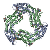
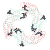
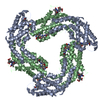
The structure of a ternary complex between CheA domains P4 and P5 with CheW and with an unzipped fragment of TM14, a chemoreceptor analog from Thermotoga maritima.
Components
Chemotaxis protein CheA
Chemotaxis protein CheW
Methyl-accepting chemotaxis protein
Keywords
IMMUNE SYSTEM / Ternary complex / Transmembrane Signaling Two component system Receptor / Histidine Kinase Adaptor protein / Membrane
Function / homology
Function and homology information
regulation of chemotaxis / phosphorelay sensor kinase activity / histidine kinase / phosphorelay signal transduction system / chemotaxis / protein domain specific binding / signal transduction / ATP binding / membrane / cytosol / cytoplasm Similarity search - Function
Single alpha-helices involved in coiled-coils or other helix-helix interfaces - #3200 / CheA-289, Domain 4 / Chemotaxis protein CheW / Chemotaxis protein CheA, P2 response regulator-binding / Chemotaxis protein CheA, P2 response regulator-binding domain superfamily / P2 response regulator binding domain / Histidine kinase CheA-like, homodimeric domain / CheY-binding domain of CheA / Histidine kinase CheA-like, homodimeric domain superfamily / Signal transducing histidine kinase, homodimeric domain ...Single alpha-helices involved in coiled-coils or other helix-helix interfaces - #3200 / CheA-289, Domain 4 / Chemotaxis protein CheW / Chemotaxis protein CheA, P2 response regulator-binding / Chemotaxis protein CheA, P2 response regulator-binding domain superfamily / P2 response regulator binding domain / Histidine kinase CheA-like, homodimeric domain / CheY-binding domain of CheA / Histidine kinase CheA-like, homodimeric domain superfamily / Signal transducing histidine kinase, homodimeric domain / Signal transducing histidine kinase, homodimeric domain / : / CheW-like domain profile. / CheW-like domain / CheW-like domain superfamily / CheW-like domain / Two component signalling adaptor domain / Histidine Phosphotransfer domain / Hpt domain / Histidine-containing phosphotransfer (HPt) domain profile. / Signal transduction histidine kinase, phosphotransfer (Hpt) domain / HPT domain superfamily / Methyl-accepting chemotaxis protein (MCP) signalling domain / Methyl-accepting chemotaxis protein (MCP) signalling domain / Bacterial chemotaxis sensory transducers domain profile. / Methyl-accepting chemotaxis-like domains (chemotaxis sensory transducer). / Signal transduction histidine kinase-related protein, C-terminal / Signal transduction histidine kinase, dimerisation/phosphoacceptor domain superfamily / SH3 Domains / Single alpha-helices involved in coiled-coils or other helix-helix interfaces / Histidine kinase domain / Histidine kinase domain profile. / Histidine kinase-, DNA gyrase B-, and HSP90-like ATPase / Helix non-globular / Histidine kinase-like ATPases / Histidine kinase/HSP90-like ATPase / Special / Histidine kinase/HSP90-like ATPase superfamily / SH3 type barrels. / OB fold (Dihydrolipoamide Acetyltransferase, E2P) / Roll / Beta Barrel / Mainly Beta Similarity search - Domain/homology
Chemotaxis protein CheA / Chemotaxis protein CheW / Methyl-accepting chemotaxis protein, putative Similarity search - Component
The receptor fragment is dimerized by crystal symmetry to produce a 4-helix bundle. The assembly state of the receptor kinase arrays is complex and extended, but the ring structure formed by the alternating CheA and CheW units built by the crystal symmetry of this structure are thought to be important features of the assembled system.
-
Components
#1: Protein
ChemotaxisproteinCheA
Mass: 35403.945 Da / Num. of mol.: 1 Source method: isolated from a genetically manipulated source Source: (gene. exp.) Thermotoga maritima (bacteria) / Strain: ATCC 43589 / MSB8 / DSM 3109 / JCM 10099 / Gene: cheA, TM_0702 / Production host: Escherichia coli (E. coli) / Strain (production host): E. coli BL-21 (RIL DE3) / References: UniProt: Q56310, histidine kinase
#2: Protein
ChemotaxisproteinCheW
Mass: 16976.834 Da / Num. of mol.: 1 Source method: isolated from a genetically manipulated source Source: (gene. exp.) Thermotoga maritima (bacteria) / Strain: ATCC 43589 / MSB8 / DSM 3109 / JCM 10099 / Gene: cheW, TM_0701 / Production host: Escherichia coli (E. coli) / Strain (production host): BL-21 (RIL DE3) / References: UniProt: Q56311
Mass: 9976.237 Da / Num. of mol.: 2 Source method: isolated from a genetically manipulated source Source: (gene. exp.) Thermotoga maritima (bacteria) / Strain: ATCC 43589 / MSB8 / DSM 3109 / JCM 10099 / Gene: ThemaDRAFT_1422, Tm00014, TM_0014 / Production host: Escherichia coli (E. coli) / Strain (production host): BL-21 (RIL DE3) / References: UniProt: Q7DFA3
-
Experimental details
-
Experiment
Experiment
Method: X-RAY DIFFRACTION / Number of used crystals: 1
-
Sample preparation
Crystal
Density Matthews: 6.34 Å3/Da / Density % sol: 80.59 %
Crystal grow
Temperature: 298 K / Method: vapor diffusion, hanging drop / pH: 8.5 Details: Cubic shaped crystals (50x50x50 um3) were grown from a mixture of 520 uM Tm14s, 457 uM CheA 354 and 121 uM CheW after 1 month by vapor diffusion from a 2 ul drop (1:1 mixture of protein and ...Details: Cubic shaped crystals (50x50x50 um3) were grown from a mixture of 520 uM Tm14s, 457 uM CheA 354 and 121 uM CheW after 1 month by vapor diffusion from a 2 ul drop (1:1 mixture of protein and reservoir: 500 ul reservoir of 0.2 M sodium acetate trihydrate, 0.1 M Tris (pH 8.5), 15% w/v polyethylene glycol 4,000), VAPOR DIFFUSION, HANGING DROP, temperature 298K
In the structure databanks used in Yorodumi, some data are registered as the other names, "COVID-19 virus" and "2019-nCoV". Here are the details of the virus and the list of structure data.
Jan 31, 2019. EMDB accession codes are about to change! (news from PDBe EMDB page)
EMDB accession codes are about to change! (news from PDBe EMDB page)
The allocation of 4 digits for EMDB accession codes will soon come to an end. Whilst these codes will remain in use, new EMDB accession codes will include an additional digit and will expand incrementally as the available range of codes is exhausted. The current 4-digit format prefixed with “EMD-” (i.e. EMD-XXXX) will advance to a 5-digit format (i.e. EMD-XXXXX), and so on. It is currently estimated that the 4-digit codes will be depleted around Spring 2019, at which point the 5-digit format will come into force.
The EM Navigator/Yorodumi systems omit the EMD- prefix.
Related info.:Q: What is EMD? / ID/Accession-code notation in Yorodumi/EM Navigator
Yorodumi is a browser for structure data from EMDB, PDB, SASBDB, etc.
This page is also the successor to EM Navigator detail page, and also detail information page/front-end page for Omokage search.
The word "yorodu" (or yorozu) is an old Japanese word meaning "ten thousand". "mi" (miru) is to see.
Related info.:EMDB / PDB / SASBDB / Comparison of 3 databanks / Yorodumi Search / Aug 31, 2016. New EM Navigator & Yorodumi / Yorodumi Papers / Jmol/JSmol / Function and homology information / Changes in new EM Navigator and Yorodumi
 Movie
Movie Controller

 Yorodumi
 Open data
 Basic information
 Components
 Keywords
 Function and homology information
 Thermotoga maritima (bacteria)
 X-RAY DIFFRACTION /
  Authors
 Citation
 Structure visualization
 Downloads & links
 Other downloads


 PDBj
PDBj


 Assembly


 Sample preparation
 / Beamline: A1 / Wavelength: 0.976 Å
 Processing