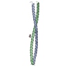
 Movie
Movie Controller

[English] 日本語
 Yorodumi
- PDB-5wlq: Crystal Structure of Amino Acids 1677-1755 of Human Beta Cardiac ... -
+ Open data

- Basic information
| Entry | Database: PDB / ID: 5wlq | ||||||
|---|---|---|---|---|---|---|---|
| Title | Crystal Structure of Amino Acids 1677-1755 of Human Beta Cardiac Myosin Fused to Gp7 and Eb1 | ||||||
 Components Components | Capsid assembly scaffolding protein,Myosin-7,Microtubule-associated protein RP/EB family member 1 | ||||||
 Keywords Keywords | MOTOR PROTEIN / Myosin / Gp7 / Coiled-Coil / Eb1 | ||||||
| Function / homology |  Function and homology information Function and homology informationviral scaffold / protein localization to astral microtubule / protein localization to mitotic spindle / cortical microtubule cytoskeleton / mitotic spindle astral microtubule end / regulation of slow-twitch skeletal muscle fiber contraction / regulation of the force of skeletal muscle contraction / protein localization to microtubule / cell projection membrane / microtubule plus-end ...viral scaffold / protein localization to astral microtubule / protein localization to mitotic spindle / cortical microtubule cytoskeleton / mitotic spindle astral microtubule end / regulation of slow-twitch skeletal muscle fiber contraction / regulation of the force of skeletal muscle contraction / protein localization to microtubule / cell projection membrane / microtubule plus-end / muscle myosin complex / mitotic spindle microtubule / transition between fast and slow fiber / cardiac muscle hypertrophy in response to stress / regulation of the force of heart contraction / attachment of mitotic spindle microtubules to kinetochore / myosin filament / microtubule plus-end binding / microtubule bundle formation / non-motile cilium assembly / muscle filament sliding / protein localization to centrosome / adult heart development / myosin complex / myosin II complex / virion assembly / ventricular cardiac muscle tissue morphogenesis / mitotic spindle pole / microfilament motor activity / spindle midzone / establishment of mitotic spindle orientation / negative regulation of microtubule polymerization / microtubule polymerization / microtubule organizing center / myofibril / regulation of microtubule polymerization or depolymerization / ATP metabolic process / positive regulation of microtubule polymerization / cytoplasmic microtubule / skeletal muscle contraction / spindle assembly / cardiac muscle contraction / stress fiber / Loss of Nlp from mitotic centrosomes / Loss of proteins required for interphase microtubule organization from the centrosome / Amplification of signal from unattached kinetochores via a MAD2 inhibitory signal / Recruitment of mitotic centrosome proteins and complexes / muscle contraction / Recruitment of NuMA to mitotic centrosomes / Anchoring of the basal body to the plasma membrane / Mitotic Prometaphase / regulation of heart rate / EML4 and NUDC in mitotic spindle formation / striated muscle contraction / AURKA Activation by TPX2 / Resolution of Sister Chromatid Cohesion / protein serine/threonine kinase binding / sarcomere / RHO GTPases Activate Formins / Z disc / actin filament binding / intracellular protein localization / The role of GTSE1 in G2/M progression after G2 checkpoint / Separation of Sister Chromatids / Regulation of PLK1 Activity at G2/M Transition / cell migration / microtubule / calmodulin binding / ciliary basal body / cadherin binding / cell division / focal adhesion / centrosome / Golgi apparatus / DNA binding / RNA binding / ATP binding / identical protein binding / cytosol / cytoplasm Similarity search - Function | ||||||
| Biological species |   Bacillus phage phi29 (virus) Bacillus phage phi29 (virus) Homo sapiens (human) Homo sapiens (human) | ||||||
| Method |  X-RAY DIFFRACTION / SYNCHROTRON / MOLECULAR REPLACEMENT / Resolution: 3.104 Å X-RAY DIFFRACTION / SYNCHROTRON / MOLECULAR REPLACEMENT / Resolution: 3.104 Å | ||||||
 Authors Authors | Andreas, M.P. / Ajay, G. / Gellings, J. / Rayment, I. | ||||||
| Funding support |  United States, 1items United States, 1items
| ||||||
 Citation Citation | Journal: J. Struct. Biol. / Year: 2017 Title: Design considerations in coiled-coil fusion constructs for the structural determination of a problematic region of the human cardiac myosin rod. Authors: Andreas, M.P. / Ajay, G. / Gellings, J.A. / Rayment, I. | ||||||
| History |
|
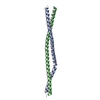
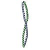
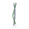
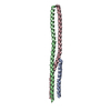
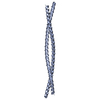
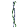
- Structure visualization
| Structure viewer | Molecule: MolmilJmol/JSmol |
|---|
- Downloads & links
-Download
| PDBx/mmCIF format | 5wlq.cif.gz | 60.1 KB | Display | PDBx/mmCIF format |
|---|---|---|---|---|
| PDB format | pdb5wlq.ent.gz | 42.2 KB | Display | PDB format |
| PDBx/mmJSON format | 5wlq.json.gz | Tree view | PDBx/mmJSON format | |
| Others |  Other downloads Other downloads |
-Validation report
| Arichive directory | https://data.pdbj.org/pub/pdb/validation_reports/wl/5wlqftp://data.pdbj.org/pub/pdb/validation_reports/wl/5wlq | HTTPS FTP |
|---|
-Related structure data
| Related structure data | 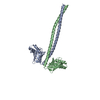 5wj7C 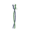 5wjbC 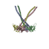 5wlzC 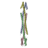 5wmeC 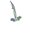 1ik9S S: Starting model for refinement C: citing same article ( |
|---|---|
| Similar structure data |

-Links
 PDBj
PDBj


- Assembly
| Deposited unit | 
| ||||||||
|---|---|---|---|---|---|---|---|---|---|
| 1 | 
| ||||||||
| Unit cell |
|
-Components
| #1: Protein | Mass: 20781.934 Da / Num. of mol.: 1 Fragment: UNP P13848 residues 2-48 UNP Q15691 residues 208-256, UNP P12883 residues 1677-1755 Source method: isolated from a genetically manipulated source Source: (gene. exp.) Bacillus phage phi29 (virus), (gene. exp.) Homo sapiens (human)Gene: MYH7, MYHCB, MAPRE1 / Production host:  References: UniProt: P13848, UniProt: P12883, UniProt: Q15691 | ||
|---|---|---|---|
| #2: Chemical | ChemComp-SO4 /   Mass: 96.063 Da / Num. of mol.: 1 / Source method: obtained synthetically / Formula: SO4 Mass: 96.063 Da / Num. of mol.: 1 / Source method: obtained synthetically / Formula: SO4 | ||
| #3: Chemical | ChemComp-TMO /   Mass: 75.110 Da / Num. of mol.: 4 / Source method: obtained synthetically / Formula: C3H9NO Mass: 75.110 Da / Num. of mol.: 4 / Source method: obtained synthetically / Formula: C3H9NO#4: Water | ChemComp-HOH / |  Mass: 18.015 Da / Num. of mol.: 8 / Source method: isolated from a natural source / Formula: H2O Mass: 18.015 Da / Num. of mol.: 8 / Source method: isolated from a natural source / Formula: H2O |
-Experimental details
-Experiment
| Experiment | Method: X-RAY DIFFRACTION / Number of used crystals: 1 |
|---|
- Sample preparation
| Crystal | Density Matthews: 6.3 Å3/Da / Density % sol: 80.4 % |
|---|---|
| Crystal grow | Temperature: 298 K / Method: vapor diffusion, hanging drop / pH: 9 Details: 1.6 M Ammonium Aulfate, 500 mM Trimethyl Ammonium N-Oxide, 100 mM Bis-tris Propane pH 9.0 |
-Data collection
| Diffraction | Mean temperature: 100 K |
|---|---|
| Diffraction source | Source: SYNCHROTRON / Site: APS / Beamline: 19-ID / Wavelength: 0.97934 Å |
| Detector | Type: ADSC QUANTUM 315r / Detector: CCD / Date: Dec 7, 2015 |
| Radiation | Protocol: SINGLE WAVELENGTH / Monochromatic (M) / Laue (L): M / Scattering type: x-ray |
| Radiation wavelength | Wavelength: 0.97934 Å / Relative weight: 1 |
| Reflection | Resolution: 3.1→50 Å / Num. obs: 9815 / % possible obs: 100 % / Redundancy: 19.6 % / Rmerge(I) obs: 0.117 / Net I/σ(I): 24.2 |
| Reflection shell | Resolution: 3.1→3.15 Å / Redundancy: 19.5 % / Rmerge(I) obs: 0.667 / Mean I/σ(I) obs: 4.46 / Num. unique obs: 478 / % possible all: 100 |
- Processing
| Software |
| |||||||||||||||||||||||||||||||||||||||||||||||||||||||||||||||||||||||||||
|---|---|---|---|---|---|---|---|---|---|---|---|---|---|---|---|---|---|---|---|---|---|---|---|---|---|---|---|---|---|---|---|---|---|---|---|---|---|---|---|---|---|---|---|---|---|---|---|---|---|---|---|---|---|---|---|---|---|---|---|---|---|---|---|---|---|---|---|---|---|---|---|---|---|---|---|---|
| Refinement | Method to determine structure: MOLECULAR REPLACEMENT Starting model: 1IK9 Resolution: 3.104→41.161 Å / SU ML: 0.29 / Cross valid method: FREE R-VALUE / σ(F): 1.35 / Phase error: 25.75
| |||||||||||||||||||||||||||||||||||||||||||||||||||||||||||||||||||||||||||
| Solvent computation | Shrinkage radii: 0.9 Å / VDW probe radii: 1.11 Å | |||||||||||||||||||||||||||||||||||||||||||||||||||||||||||||||||||||||||||
| Refinement step | Cycle: LAST / Resolution: 3.104→41.161 Å
| |||||||||||||||||||||||||||||||||||||||||||||||||||||||||||||||||||||||||||
| Refine LS restraints |
| |||||||||||||||||||||||||||||||||||||||||||||||||||||||||||||||||||||||||||
| LS refinement shell |
| |||||||||||||||||||||||||||||||||||||||||||||||||||||||||||||||||||||||||||
| Refinement TLS params. | Method: refined / Refine-ID: X-RAY DIFFRACTION
| |||||||||||||||||||||||||||||||||||||||||||||||||||||||||||||||||||||||||||
| Refinement TLS group |
|
