 ムービー
ムービー コントローラー


+ データを開く

- 基本情報
基本情報
| 登録情報 | データベース: PDB / ID: 6ylu | ||||||
|---|---|---|---|---|---|---|---|
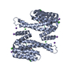
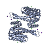
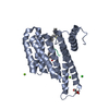
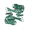
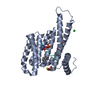
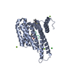
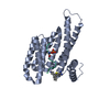
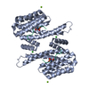
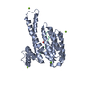
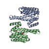
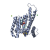
| タイトル | 14-3-3sigma in complex with BLNKpT152 phosphopeptide crystal structure | ||||||
 要素 要素 |
| ||||||
 キーワード キーワード | PEPTIDE BINDING PROTEIN / Adaptor Protein / Phosphorylation | ||||||
| 機能・相同性 |  機能・相同性情報 機能・相同性情報transmembrane receptor protein tyrosine kinase adaptor activity / regulation of epidermal cell division / protein kinase C inhibitor activity / positive regulation of epidermal cell differentiation / keratinocyte development / keratinization / regulation of cell-cell adhesion / humoral immune response / cAMP/PKA signal transduction / Regulation of localization of FOXO transcription factors ...transmembrane receptor protein tyrosine kinase adaptor activity / regulation of epidermal cell division / protein kinase C inhibitor activity / positive regulation of epidermal cell differentiation / keratinocyte development / keratinization / regulation of cell-cell adhesion / humoral immune response / cAMP/PKA signal transduction / Regulation of localization of FOXO transcription factors / keratinocyte proliferation / phosphoserine residue binding / Activation of BAD and translocation to mitochondria / negative regulation of keratinocyte proliferation / establishment of skin barrier / negative regulation of protein localization to plasma membrane / Chk1/Chk2(Cds1) mediated inactivation of Cyclin B:Cdk1 complex / SARS-CoV-2 targets host intracellular signalling and regulatory pathways / negative regulation of protein kinase activity / negative regulation of stem cell proliferation / phospholipase binding / SARS-CoV-1 targets host intracellular signalling and regulatory pathways / RHO GTPases activate PKNs / positive regulation of protein localization / signaling adaptor activity / SH2 domain binding / positive regulation of cell adhesion / protein sequestering activity / protein export from nucleus / negative regulation of innate immune response / cell surface receptor protein tyrosine kinase signaling pathway / protein tyrosine kinase binding / Antigen activates B Cell Receptor (BCR) leading to generation of second messengers / TP53 Regulates Transcription of Genes Involved in G2 Cell Cycle Arrest / release of cytochrome c from mitochondria / B cell differentiation / positive regulation of protein export from nucleus / stem cell proliferation / B cell receptor signaling pathway / TP53 Regulates Metabolic Genes / Translocation of SLC2A4 (GLUT4) to the plasma membrane / Regulation of signaling by CBL / molecular condensate scaffold activity / intrinsic apoptotic signaling pathway in response to DNA damage / intracellular protein localization / regulation of protein localization / positive regulation of cell growth / Potential therapeutics for SARS / regulation of cell cycle / intracellular signal transduction / cadherin binding / inflammatory response / intracellular membrane-bounded organelle / lipid binding / positive regulation of gene expression / protein kinase binding / enzyme binding / negative regulation of transcription by RNA polymerase II / signal transduction / extracellular space / extracellular exosome / identical protein binding / nucleus / membrane / plasma membrane / cytoplasm / cytosol 類似検索 - 分子機能 | ||||||
| 生物種 |  Homo sapiens (ヒト) Homo sapiens (ヒト) | ||||||
| 手法 |  X線回折 / 分子置換 / 解像度: 1.88 Å X線回折 / 分子置換 / 解像度: 1.88 Å | ||||||
 データ登録者 データ登録者 | Soini, L. / Leysen, S. / Davis, J. / Ottmann, C. | ||||||
| 資金援助 |  オランダ, 1件 オランダ, 1件
| ||||||
 引用 引用 | ジャーナル: J.Struct.Biol. / 年: 2020 タイトル: 14-3-3sigma in complex with BLNKpT152 phosphopeptide crystal structure 著者: Soini, L. / Leysen, S. / Davis, J. / Ottmann, C. | ||||||
| 履歴 |
|
- 構造の表示
構造の表示
| 構造ビューア | 分子: MolmilJmol/JSmol |
|---|
- ダウンロードとリンク
-ダウンロード
| PDBx/mmCIF形式 | 6ylu.cif.gz | 91 KB | 表示 | PDBx/mmCIF形式 |
|---|---|---|---|---|
| PDB形式 | pdb6ylu.ent.gz | 54.5 KB | 表示 | PDB形式 |
| PDBx/mmJSON形式 | 6ylu.json.gz | ツリー表示 | PDBx/mmJSON形式 | |
| その他 |  その他のダウンロード その他のダウンロード |
-検証レポート
| 文書・要旨 | 6ylu_validation.pdf.gz | 432.9 KB | 表示 | wwPDB検証レポート |
|---|---|---|---|---|
| 文書・詳細版 | 6ylu_full_validation.pdf.gz | 433.5 KB | 表示 | |
| XML形式データ | 6ylu_validation.xml.gz | 15.5 KB | 表示 | |
| CIF形式データ | 6ylu_validation.cif.gz | 24.4 KB | 表示 | |
| アーカイブディレクトリ | https://data.pdbj.org/pub/pdb/validation_reports/yl/6yluftp://data.pdbj.org/pub/pdb/validation_reports/yl/6ylu | HTTPS FTP |
-関連構造データ
| 関連構造データ | 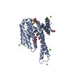 3mhrS S: 精密化の開始モデル |
|---|---|
| 類似構造データ |


-リンク
 PDBj
PDBj


- 集合体
集合体
| 登録構造単位 | 
| ||||||||||||
|---|---|---|---|---|---|---|---|---|---|---|---|---|---|
| 1 | 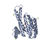
| ||||||||||||
| 単位格子 |
| ||||||||||||
| Components on special symmetry positions |
|
-要素
| #1: タンパク質 | 分子量: 26542.914 Da / 分子数: 1 / 由来タイプ: 組換発現 / 由来: (組換発現) Homo sapiens (ヒト) / 遺伝子: SFN, HME1 / 発現宿主:  | ||||||
|---|---|---|---|---|---|---|---|
| #2: タンパク質・ペプチド | 分子量: 1295.400 Da / 分子数: 1 / 由来タイプ: 合成 / 由来: (合成) Homo sapiens (ヒト) / 参照: UniProt: Q8WV28*PLUS | ||||||
| #3: 化合物 |   分子量: 24.305 Da / 分子数: 3 / 由来タイプ: 合成 / 式: Mg 分子量: 24.305 Da / 分子数: 3 / 由来タイプ: 合成 / 式: Mg#4: 水 | ChemComp-HOH / |  分子量: 18.015 Da / 分子数: 387 / 由来タイプ: 天然 / 式: H2O 分子量: 18.015 Da / 分子数: 387 / 由来タイプ: 天然 / 式: H2O研究の焦点であるリガンドがあるか | Y | Has protein modification | Y | |
-実験情報
-実験
| 実験 | 手法: X線回折 / 使用した結晶の数: 1 |
|---|
- 試料調製
試料調製
| 結晶 | マシュー密度: 2.72 Å3/Da / 溶媒含有率: 54.8 % |
|---|---|
| 結晶化 | 温度: 277.15 K / 手法: 蒸気拡散法, シッティングドロップ法 / pH: 7.5 詳細: 95 mM Hepes pH 7.1-7.7, 24-29% PEG400, 190 mM CaCl2, Glycerol 5% PH範囲: 7.1-7-7 |
-データ収集
| 回折 | 平均測定温度: 80 K / Serial crystal experiment: N |
|---|---|
| 放射光源 | 由来: SEALED TUBE / タイプ: RIGAKU MICROMAX-003 / 波長: 1.541 Å |
| 検出器 | タイプ: DECTRIS PILATUS3 R 200K-A / 検出器: PIXEL / 日付: 2016年11月14日 |
| 放射 | プロトコル: SINGLE WAVELENGTH / 単色(M)・ラウエ(L): M / 散乱光タイプ: x-ray |
| 放射波長 | 波長: 1.541 Å / 相対比: 1 |
| 反射 | 解像度: 1.88→26.63 Å / Num. obs: 23758 / % possible obs: 99.9 % / 冗長度: 5.1 % / Biso Wilson estimate: 11.73 Å2 / CC1/2: 0.99 / Rmerge(I) obs: 0.123 / Rpim(I) all: 0.089 / Rrim(I) all: 0.152 / Net I/σ(I): 8.8 |
| 反射 シェル | 解像度: 1.88→1.93 Å / Num. unique obs: 1473 / CC1/2: 0.915 |
- 解析
解析
| ソフトウェア |
| |||||||||||||||||||||||||||||||||||||||||||||||||||||||||||||||
|---|---|---|---|---|---|---|---|---|---|---|---|---|---|---|---|---|---|---|---|---|---|---|---|---|---|---|---|---|---|---|---|---|---|---|---|---|---|---|---|---|---|---|---|---|---|---|---|---|---|---|---|---|---|---|---|---|---|---|---|---|---|---|---|---|
| 精密化 | 構造決定の手法: 分子置換 開始モデル: 3MHR 解像度: 1.88→26.63 Å / SU ML: 0.1692 / 交差検証法: FREE R-VALUE / σ(F): 1.34 / 位相誤差: 19.393
| |||||||||||||||||||||||||||||||||||||||||||||||||||||||||||||||
| 溶媒の処理 | 減衰半径: 0.9 Å / VDWプローブ半径: 1.11 Å | |||||||||||||||||||||||||||||||||||||||||||||||||||||||||||||||
| 原子変位パラメータ | Biso mean: 15.57 Å2 | |||||||||||||||||||||||||||||||||||||||||||||||||||||||||||||||
| 精密化ステップ | サイクル: LAST / 解像度: 1.88→26.63 Å
| |||||||||||||||||||||||||||||||||||||||||||||||||||||||||||||||
| 拘束条件 |
| |||||||||||||||||||||||||||||||||||||||||||||||||||||||||||||||
| LS精密化 シェル |
|
