 Movie
Movie Controller

[English] 日本語
 Yorodumi
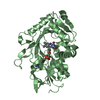
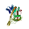
- PDB-6y8o: Mycobacterium smegmatis GyrB 22kDa ATPase sub-domain in complex w... -
+ Open data

- Basic information
| Entry | Database: PDB / ID: 6y8o | |||||||||
|---|---|---|---|---|---|---|---|---|---|---|
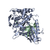
| Title | Mycobacterium smegmatis GyrB 22kDa ATPase sub-domain in complex with novobiocin | |||||||||
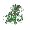
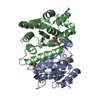
 Components Components | DNA gyrase subunit B | |||||||||
 Keywords Keywords | DNA BINDING PROTEIN / Binding Sites / DNA Gyrase / inhibitors / / novobiocin IV / ISOMERASE | |||||||||
| Function / homology |  Function and homology information Function and homology informationDNA negative supercoiling activity / DNA topoisomerase (ATP-hydrolysing) / DNA topological change / DNA-templated DNA replication / chromosome / DNA binding / ATP binding / metal ion binding / cytoplasm Similarity search - Function | |||||||||
| Biological species |  Mycolicibacterium smegmatis (bacteria) Mycolicibacterium smegmatis (bacteria) | |||||||||
| Method |  X-RAY DIFFRACTION / SYNCHROTRON / MOLECULAR REPLACEMENT / Resolution: 1.6 Å X-RAY DIFFRACTION / SYNCHROTRON / MOLECULAR REPLACEMENT / Resolution: 1.6 Å | |||||||||
 Authors Authors | Henderson, S.R. / Stevenson, C.E.M. / Malone, B. / Zholnerovych, Y. / Mitchenall, L.A. / Pichowicz, M. / McGarry, D.H. / Cooper, I.R. / Charrier, C. / Salisbury, A. ...Henderson, S.R. / Stevenson, C.E.M. / Malone, B. / Zholnerovych, Y. / Mitchenall, L.A. / Pichowicz, M. / McGarry, D.H. / Cooper, I.R. / Charrier, C. / Salisbury, A. / Lawson, D.M. / Maxwell, A. | |||||||||
| Funding support |  United Kingdom, 2items United Kingdom, 2items
| |||||||||
 Citation Citation | Journal: J.Antimicrob.Chemother. / Year: 2020 Title: Structural and mechanistic analysis of ATPase inhibitors targeting mycobacterial DNA gyrase. Authors: Henderson, S.R. / Stevenson, C.E.M. / Malone, B. / Zholnerovych, Y. / Mitchenall, L.A. / Pichowicz, M. / McGarry, D.H. / Cooper, I.R. / Charrier, C. / Salisbury, A.M. / Lawson, D.M. / Maxwell, A. | |||||||||
| History |
|
- Structure visualization
| Structure viewer | Molecule: MolmilJmol/JSmol |
|---|
- Downloads & links
-Download
| PDBx/mmCIF format | 6y8o.cif.gz | 196.3 KB | Display | PDBx/mmCIF format |
|---|---|---|---|---|
| PDB format | pdb6y8o.ent.gz | 154.1 KB | Display | PDB format |
| PDBx/mmJSON format | 6y8o.json.gz | Tree view | PDBx/mmJSON format | |
| Others |  Other downloads Other downloads |
-Validation report
| Arichive directory | https://data.pdbj.org/pub/pdb/validation_reports/y8/6y8oftp://data.pdbj.org/pub/pdb/validation_reports/y8/6y8o | HTTPS FTP |
|---|
-Related structure data
| Related structure data |  6y8lC  6y8nC  4b6cS C: citing same article ( S: Starting model for refinement |
|---|---|
| Similar structure data |


-Links
 PDBj
PDBj


- Assembly
| Deposited unit | 
| ||||||||||||||||||
|---|---|---|---|---|---|---|---|---|---|---|---|---|---|---|---|---|---|---|---|
| 1 |
| ||||||||||||||||||
| Unit cell |
| ||||||||||||||||||
| Noncrystallographic symmetry (NCS) | NCS domain:
NCS domain segments: Component-ID: _ / Ens-ID: 1 / Beg auth comp-ID: THR / Beg label comp-ID: THR / End auth comp-ID: PRO / End label comp-ID: PRO / Refine code: _ / Auth seq-ID: 18 - 254 / Label seq-ID: 29 - 233
|
-Components
-Protein , 1 types, 2 molecules AB
| #1: Protein | Mass: 26152.248 Da / Num. of mol.: 2 Source method: isolated from a genetically manipulated source Source: (gene. exp.) Mycolicibacterium smegmatis (bacteria) / Gene: gyrB / Production host: References: UniProt: P0C559, UniProt: A0QNE0*PLUS, DNA topoisomerase (ATP-hydrolysing) |
|---|
-Non-polymers , 5 types, 398 molecules 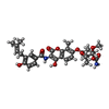


| #2: Chemical |  Mass: 612.624 Da / Num. of mol.: 2 / Source method: obtained synthetically / Formula: C31H36N2O11 / Feature type: SUBJECT OF INVESTIGATION / Comment: antibiotic*YM Mass: 612.624 Da / Num. of mol.: 2 / Source method: obtained synthetically / Formula: C31H36N2O11 / Feature type: SUBJECT OF INVESTIGATION / Comment: antibiotic*YM#3: Chemical | ChemComp-EDO /  Mass: 62.068 Da / Num. of mol.: 10 / Source method: obtained synthetically / Formula: C2H6O2 Mass: 62.068 Da / Num. of mol.: 10 / Source method: obtained synthetically / Formula: C2H6O2#4: Chemical |  Mass: 59.044 Da / Num. of mol.: 3 / Source method: obtained synthetically / Formula: C2H3O2 Mass: 59.044 Da / Num. of mol.: 3 / Source method: obtained synthetically / Formula: C2H3O2#5: Chemical | ChemComp-NA /  Mass: 22.990 Da / Num. of mol.: 5 / Source method: obtained synthetically / Formula: Na Mass: 22.990 Da / Num. of mol.: 5 / Source method: obtained synthetically / Formula: Na#6: Water | ChemComp-HOH / | Mass: 18.015 Da / Num. of mol.: 378 / Source method: isolated from a natural source / Formula: H2O |
|---|
-Details
| Has ligand of interest | Y |
|---|
-Experimental details
-Experiment
| Experiment | Method: X-RAY DIFFRACTION / Number of used crystals: 1 |
|---|
- Sample preparation
| Crystal | Density Matthews: 2.31 Å3/Da / Density % sol: 46.66 % |
|---|---|
| Crystal grow | Temperature: 293 K / Method: vapor diffusion, sitting drop / pH: 5.6 Details: Crystals grown with 17 mg/ml protein in the presence of 1 mM novobiocin against 15% (w/v) PEG8000, 100 mM sodium acetate pH 5.6, 200 mM calcium acetate were harvested in the precipitant ...Details: Crystals grown with 17 mg/ml protein in the presence of 1 mM novobiocin against 15% (w/v) PEG8000, 100 mM sodium acetate pH 5.6, 200 mM calcium acetate were harvested in the precipitant supplemented with 25% (v/v) ethylene glycol for cryoprotection. |
-Data collection
| Diffraction | Mean temperature: 100 K / Serial crystal experiment: N | ||||||||||||||||||||||||||||||
|---|---|---|---|---|---|---|---|---|---|---|---|---|---|---|---|---|---|---|---|---|---|---|---|---|---|---|---|---|---|---|---|
| Diffraction source | Source: SYNCHROTRON / Site: Diamond / Beamline: I04-1 / Wavelength: 0.9159 Å | ||||||||||||||||||||||||||||||
| Detector | Type: DECTRIS PILATUS 6M / Detector: PIXEL / Date: Mar 9, 2018 | ||||||||||||||||||||||||||||||
| Radiation | Protocol: SINGLE WAVELENGTH / Monochromatic (M) / Laue (L): M / Scattering type: x-ray | ||||||||||||||||||||||||||||||
| Radiation wavelength | Wavelength: 0.9159 Å / Relative weight: 1 | ||||||||||||||||||||||||||||||
| Reflection | Resolution: 1.6→78.48 Å / Num. obs: 58273 / % possible obs: 100 % / Redundancy: 6.6 % / CC1/2: 0.999 / Rmerge(I) obs: 0.11 / Rpim(I) all: 0.046 / Rrim(I) all: 0.119 / Net I/σ(I): 10.4 / Num. measured all: 387422 / Scaling rejects: 148 | ||||||||||||||||||||||||||||||
| Reflection shell | Diffraction-ID: 1
|
- Processing
| Software |
| |||||||||||||||||||||||||||||||||||||||||||||||||||||||||||||||||
|---|---|---|---|---|---|---|---|---|---|---|---|---|---|---|---|---|---|---|---|---|---|---|---|---|---|---|---|---|---|---|---|---|---|---|---|---|---|---|---|---|---|---|---|---|---|---|---|---|---|---|---|---|---|---|---|---|---|---|---|---|---|---|---|---|---|---|
| Refinement | Method to determine structure: MOLECULAR REPLACEMENT Starting model: 4B6C Resolution: 1.6→78.48 Å / Cor.coef. Fo:Fc: 0.979 / Cor.coef. Fo:Fc free: 0.965 / SU B: 4.628 / SU ML: 0.067 / SU R Cruickshank DPI: 0.0873 / Cross valid method: THROUGHOUT / σ(F): 0 / ESU R: 0.087 / ESU R Free: 0.078 Details: HYDROGENS HAVE BEEN ADDED IN THE RIDING POSITIONS U VALUES : REFINED INDIVIDUALLY
| |||||||||||||||||||||||||||||||||||||||||||||||||||||||||||||||||
| Solvent computation | Ion probe radii: 0.8 Å / Shrinkage radii: 0.8 Å / VDW probe radii: 1.2 Å | |||||||||||||||||||||||||||||||||||||||||||||||||||||||||||||||||
| Displacement parameters | Biso max: 81.5 Å2 / Biso mean: 21.903 Å2 / Biso min: 9.86 Å2
| |||||||||||||||||||||||||||||||||||||||||||||||||||||||||||||||||
| Refinement step | Cycle: final / Resolution: 1.6→78.48 Å
| |||||||||||||||||||||||||||||||||||||||||||||||||||||||||||||||||
| Refine LS restraints |
| |||||||||||||||||||||||||||||||||||||||||||||||||||||||||||||||||
| Refine LS restraints NCS | Ens-ID: 1 / Number: 6058 / Refine-ID: X-RAY DIFFRACTION / Type: interatomic distance / Rms dev position: 0.11 Å / Weight position: 0.05
| |||||||||||||||||||||||||||||||||||||||||||||||||||||||||||||||||
| LS refinement shell | Resolution: 1.6→1.642 Å / Rfactor Rfree error: 0 / Total num. of bins used: 20
|
