 Movie
Movie Controller

[English] 日本語
 Yorodumi
- PDB-6y4p: Calmodulin N53I variant bound to cardiac ryanodine receptor (RyR2... -
+ Open data

- Basic information
| Entry | Database: PDB / ID: 6y4p | |||||||||||||||
|---|---|---|---|---|---|---|---|---|---|---|---|---|---|---|---|---|
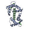
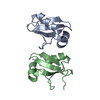
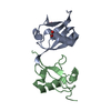
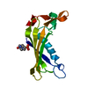
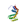
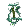
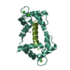
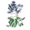
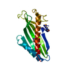
| Title | Calmodulin N53I variant bound to cardiac ryanodine receptor (RyR2) calmodulin binding domain | |||||||||||||||
 Components Components |
| |||||||||||||||
 Keywords Keywords | MEMBRANE PROTEIN / ion channel / calcium / calmodulin | |||||||||||||||
| Function / homology |  Function and homology information Function and homology informationjunctional sarcoplasmic reticulum membrane / sarcoplasmic reticulum calcium ion transport / establishment of protein localization to endoplasmic reticulum / type B pancreatic cell apoptotic process / Purkinje myocyte to ventricular cardiac muscle cell signaling / calcium-induced calcium release activity / regulation of atrial cardiac muscle cell action potential / left ventricular cardiac muscle tissue morphogenesis / suramin binding / regulation of AV node cell action potential ...junctional sarcoplasmic reticulum membrane / sarcoplasmic reticulum calcium ion transport / establishment of protein localization to endoplasmic reticulum / type B pancreatic cell apoptotic process / Purkinje myocyte to ventricular cardiac muscle cell signaling / calcium-induced calcium release activity / regulation of atrial cardiac muscle cell action potential / left ventricular cardiac muscle tissue morphogenesis / suramin binding / regulation of AV node cell action potential / regulation of SA node cell action potential / Stimuli-sensing channels / cell communication by electrical coupling involved in cardiac conduction / regulation of ventricular cardiac muscle cell action potential / ventricular cardiac muscle cell action potential / positive regulation of sequestering of calcium ion / Ion homeostasis / embryonic heart tube morphogenesis / cardiac muscle hypertrophy / calcium ion transport into cytosol / ryanodine-sensitive calcium-release channel activity / CaM pathway / Cam-PDE 1 activation / Sodium/Calcium exchangers / response to caffeine / regulation of cardiac muscle contraction by calcium ion signaling / release of sequestered calcium ion into cytosol by sarcoplasmic reticulum / Calmodulin induced events / response to redox state / Reduction of cytosolic Ca++ levels / Activation of Ca-permeable Kainate Receptor / CREB1 phosphorylation through the activation of CaMKII/CaMKK/CaMKIV cascasde / Loss of phosphorylation of MECP2 at T308 / CREB1 phosphorylation through the activation of Adenylate Cyclase / CaMK IV-mediated phosphorylation of CREB / PKA activation / negative regulation of high voltage-gated calcium channel activity / calcium ion transmembrane import into cytosol / Glycogen breakdown (glycogenolysis) / CLEC7A (Dectin-1) induces NFAT activation / Activation of RAC1 downstream of NMDARs / negative regulation of ryanodine-sensitive calcium-release channel activity / organelle localization by membrane tethering / mitochondrion-endoplasmic reticulum membrane tethering / autophagosome membrane docking / negative regulation of calcium ion export across plasma membrane / regulation of cardiac muscle cell action potential / presynaptic endocytosis / protein kinase A regulatory subunit binding / response to muscle activity / Synthesis of IP3 and IP4 in the cytosol / regulation of cell communication by electrical coupling involved in cardiac conduction / protein kinase A catalytic subunit binding / Phase 0 - rapid depolarisation / calcineurin-mediated signaling / Negative regulation of NMDA receptor-mediated neuronal transmission / positive regulation of the force of heart contraction / Unblocking of NMDA receptors, glutamate binding and activation / cellular response to caffeine / RHO GTPases activate PAKs / intracellularly gated calcium channel activity / Ion transport by P-type ATPases / Uptake and function of anthrax toxins / regulation of ryanodine-sensitive calcium-release channel activity / Long-term potentiation / protein phosphatase activator activity / Calcineurin activates NFAT / Regulation of MECP2 expression and activity / DARPP-32 events / catalytic complex / Smooth Muscle Contraction / detection of calcium ion / regulation of cardiac muscle contraction / regulation of cytosolic calcium ion concentration / RHO GTPases activate IQGAPs / regulation of cardiac muscle contraction by regulation of the release of sequestered calcium ion / smooth endoplasmic reticulum / positive regulation of heart rate / cellular response to interferon-beta / Protein methylation / calcium channel inhibitor activity / presynaptic cytosol / Activation of AMPK downstream of NMDARs / cardiac muscle contraction / Ion homeostasis / regulation of release of sequestered calcium ion into cytosol by sarcoplasmic reticulum / eNOS activation / titin binding / Tetrahydrobiopterin (BH4) synthesis, recycling, salvage and regulation / sperm midpiece / regulation of calcium-mediated signaling / response to muscle stretch / release of sequestered calcium ion into cytosol / cellular response to epinephrine stimulus / voltage-gated potassium channel complex / calcium channel complex / substantia nigra development / FCERI mediated Ca+2 mobilization / sarcoplasmic reticulum membrane / Ras activation upon Ca2+ influx through NMDA receptor Similarity search - Function | |||||||||||||||
| Biological species |  Homo sapiens (human) Homo sapiens (human) | |||||||||||||||
| Method |  X-RAY DIFFRACTION / SYNCHROTRON / MOLECULAR REPLACEMENT / Resolution: 2.13325578805 Å X-RAY DIFFRACTION / SYNCHROTRON / MOLECULAR REPLACEMENT / Resolution: 2.13325578805 Å | |||||||||||||||
 Authors Authors | Lau, K. / Nielsen, L.H. / Holt, C. / Brohus, M. / Sorensen, A.B. / Larsen, K.T. / Sommer, C. / Van Petegem, F. / Overgaard, M.T. / Wimmer, R. | |||||||||||||||
| Funding support |  Denmark, Denmark,  Germany, Germany,  Canada, 4items Canada, 4items
| |||||||||||||||
 Citation Citation | Journal: J.Biol.Chem. / Year: 2020 Title: The arrhythmogenic N53I variant subtly changes the structure and dynamics in the calmodulin N-terminal domain, altering its interaction with the cardiac ryanodine receptor. Authors: Holt, C. / Hamborg, L. / Lau, K. / Brohus, M. / Sorensen, A.B. / Larsen, K.T. / Sommer, C. / Van Petegem, F. / Overgaard, M.T. / Wimmer, R. | |||||||||||||||
| History |
|
- Structure visualization
| Structure viewer | Molecule: MolmilJmol/JSmol |
|---|
- Downloads & links
-Download
| PDBx/mmCIF format | 6y4p.cif.gz | 57.7 KB | Display | PDBx/mmCIF format |
|---|---|---|---|---|
| PDB format | pdb6y4p.ent.gz | 32.2 KB | Display | PDB format |
| PDBx/mmJSON format | 6y4p.json.gz | Tree view | PDBx/mmJSON format | |
| Others |  Other downloads Other downloads |
-Validation report
| Summary document | 6y4p_validation.pdf.gz | 1.9 MB | Display | wwPDB validaton report |
|---|---|---|---|---|
| Full document | 6y4p_full_validation.pdf.gz | 1.9 MB | Display | |
| Data in XML | 6y4p_validation.xml.gz | 8.6 KB | Display | |
| Data in CIF | 6y4p_validation.cif.gz | 11.2 KB | Display | |
| Arichive directory | https://data.pdbj.org/pub/pdb/validation_reports/y4/6y4pftp://data.pdbj.org/pub/pdb/validation_reports/y4/6y4p | HTTPS FTP |
-Related structure data
| Related structure data |  6y4oC 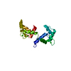 6y94C 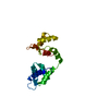 6y95C  2bcxS S: Starting model for refinement C: citing same article ( |
|---|---|
| Similar structure data |

-Links
 PDBj
PDBj


- Assembly
| Deposited unit | 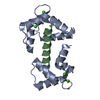
| ||||||||||||
|---|---|---|---|---|---|---|---|---|---|---|---|---|---|
| 1 |
| ||||||||||||
| Unit cell |
|
-Components
| #1: Protein | Mass: 16851.600 Da / Num. of mol.: 1 / Mutation: N53I Source method: isolated from a genetically manipulated source Source: (gene. exp.) Homo sapiens (human) / Gene: CALM1, CALM, CAM, CAM1 / Production host:  | ||||
|---|---|---|---|---|---|
| #2: Protein/peptide | Mass: 3478.235 Da / Num. of mol.: 1 Source method: isolated from a genetically manipulated source Source: (gene. exp.) | ||||
| #3: Chemical | ChemComp-CA /   Mass: 40.078 Da / Num. of mol.: 4 / Source method: obtained synthetically / Formula: Ca / Feature type: SUBJECT OF INVESTIGATION Mass: 40.078 Da / Num. of mol.: 4 / Source method: obtained synthetically / Formula: Ca / Feature type: SUBJECT OF INVESTIGATION#4: Water | ChemComp-HOH / |  Mass: 18.015 Da / Num. of mol.: 61 / Source method: isolated from a natural source / Formula: H2O Mass: 18.015 Da / Num. of mol.: 61 / Source method: isolated from a natural source / Formula: H2OHas ligand of interest | Y | |
-Experimental details
-Experiment
| Experiment | Method: X-RAY DIFFRACTION / Number of used crystals: 1 |
|---|
- Sample preparation
| Crystal grow | Temperature: 298.15 K / Method: vapor diffusion, hanging drop / Details: 0.1 M Sodium Acetate pH 4.70 and 23 % PEG 550 MME |
|---|
-Data collection
| Diffraction | Mean temperature: 100 K / Serial crystal experiment: N |
|---|---|
| Diffraction source | Source: SYNCHROTRON / Site: CLSI / Beamline: 08B1-1 / Wavelength: 1 Å |
| Detector | Type: RAYONIX MX-225 / Detector: CCD / Date: Dec 11, 2014 |
| Radiation | Protocol: SINGLE WAVELENGTH / Monochromatic (M) / Laue (L): M / Scattering type: x-ray |
| Radiation wavelength | Wavelength: 1 Å / Relative weight: 1 |
| Reflection | Resolution: 2.133→45.18 Å / Num. obs: 8489 / % possible obs: 99.76 % / Redundancy: 5.9 % / Biso Wilson estimate: 24.3986621334 Å2 / CC1/2: 0.993 / Net I/σ(I): 9.45 |
| Reflection shell | Resolution: 2.133→2.209 Å / Num. unique obs: 831 / CC1/2: 0.946 |
- Processing
| Software |
| |||||||||||||||||||||||||||||||||||||||||||||||||
|---|---|---|---|---|---|---|---|---|---|---|---|---|---|---|---|---|---|---|---|---|---|---|---|---|---|---|---|---|---|---|---|---|---|---|---|---|---|---|---|---|---|---|---|---|---|---|---|---|---|---|
| Refinement | Method to determine structure: MOLECULAR REPLACEMENT Starting model: 2bcx Resolution: 2.13325578805→45.1799 Å / SU ML: 0.232891903909 / Cross valid method: FREE R-VALUE / σ(F): 1.43770529883 / Phase error: 21.642731085
| |||||||||||||||||||||||||||||||||||||||||||||||||
| Solvent computation | Shrinkage radii: 0.9 Å / VDW probe radii: 1.11 Å | |||||||||||||||||||||||||||||||||||||||||||||||||
| Displacement parameters | Biso mean: 29.4527987444 Å2 | |||||||||||||||||||||||||||||||||||||||||||||||||
| Refinement step | Cycle: LAST / Resolution: 2.13325578805→45.1799 Å
| |||||||||||||||||||||||||||||||||||||||||||||||||
| Refine LS restraints |
| |||||||||||||||||||||||||||||||||||||||||||||||||
| LS refinement shell |
|
