 Movie
Movie Controller

[English] 日本語
 Yorodumi
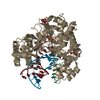
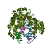
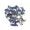
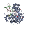
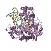
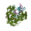
- PDB-1bvq: THREE-DIMENSIONAL STRUCTURE OF 4-HYDROXYBENZOYL COA THIOESTERASE ... -
+ Open data

- Basic information
| Entry | Database: PDB / ID: 1bvq | ||||||
|---|---|---|---|---|---|---|---|
| Title | THREE-DIMENSIONAL STRUCTURE OF 4-HYDROXYBENZOYL COA THIOESTERASE FROM PSEUDOMONAS SP. STRAIN CBS-3. | ||||||
 Components Components | PROTEIN (4-HYDROXYBENZOYL COA THIOESTERASE) | ||||||
 Keywords Keywords | HYDROLASE | ||||||
| Function / homology |  Function and homology information Function and homology information4-hydroxybenzoyl-CoA thioesterase / 4-hydroxybenzoyl-CoA thioesterase activity Similarity search - Function | ||||||
| Biological species |  Pseudomonas sp. CBS3 (bacteria) Pseudomonas sp. CBS3 (bacteria) | ||||||
| Method |  X-RAY DIFFRACTION / MIR / Resolution: 2 Å X-RAY DIFFRACTION / MIR / Resolution: 2 Å | ||||||
 Authors Authors | Holden, H.M. / Benning, M.M. / Dunaway-Mariano, D. | ||||||
 Citation Citation | Journal: J.Biol.Chem. / Year: 1998 Title: The three-dimensional structure of 4-hydroxybenzoyl-CoA thioesterase from Pseudomonas sp. Strain CBS-3. Authors: Benning, M.M. / Wesenberg, G. / Liu, R. / Taylor, K.L. / Dunaway-Mariano, D. / Holden, H.M. | ||||||
| History |
|
- Structure visualization
| Structure viewer | Molecule: MolmilJmol/JSmol |
|---|
- Downloads & links
-Download
| PDBx/mmCIF format | 1bvq.cif.gz | 41.4 KB | Display | PDBx/mmCIF format |
|---|---|---|---|---|
| PDB format | pdb1bvq.ent.gz | 28.9 KB | Display | PDB format |
| PDBx/mmJSON format | 1bvq.json.gz | Tree view | PDBx/mmJSON format | |
| Others |  Other downloads Other downloads |
-Validation report
| Arichive directory | https://data.pdbj.org/pub/pdb/validation_reports/bv/1bvqftp://data.pdbj.org/pub/pdb/validation_reports/bv/1bvq | HTTPS FTP |
|---|
-Related structure data
| Similar structure data |
|---|


-Links
 PDBj
PDBj
- Assembly
| Deposited unit | 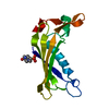
| ||||||||
|---|---|---|---|---|---|---|---|---|---|
| 1 | 
| ||||||||
| Unit cell |
|
-Components
| #1: Protein | Mass: 16140.452 Da / Num. of mol.: 1 / Source method: isolated from a natural source / Source: (natural) Pseudomonas sp. CBS3 (bacteria) / Strain: CBS-3 / References: UniProt: P56653, EC: 3.8.1.6 |
|---|---|
| #2: Chemical | ChemComp-EPE /   Mass: 238.305 Da / Num. of mol.: 1 / Source method: obtained synthetically / Formula: C8H18N2O4S / Comment: pH buffer*YM Mass: 238.305 Da / Num. of mol.: 1 / Source method: obtained synthetically / Formula: C8H18N2O4S / Comment: pH buffer*YM |
| #3: Water | ChemComp-HOH /  Mass: 18.015 Da / Num. of mol.: 66 / Source method: isolated from a natural source / Formula: H2O Mass: 18.015 Da / Num. of mol.: 66 / Source method: isolated from a natural source / Formula: H2O |
-Experimental details
-Experiment
| Experiment | Method: X-RAY DIFFRACTION / Number of used crystals: 1 |
|---|
- Sample preparation
| Crystal | Density Matthews: 2.03 Å3/Da / Density % sol: 39 % | ||||||||||||||||||||||||||||||
|---|---|---|---|---|---|---|---|---|---|---|---|---|---|---|---|---|---|---|---|---|---|---|---|---|---|---|---|---|---|---|---|
| Crystal grow | pH: 7 Details: 0.9 M AMMONIUM SULFATE 1.0 % 2-METHYL-2,4-PENTADIOL 50 MM HEPES PH 7.0 | ||||||||||||||||||||||||||||||
| Crystal | *PLUS | ||||||||||||||||||||||||||||||
| Crystal grow | *PLUS Temperature: 4 ℃ / Method: batch method | ||||||||||||||||||||||||||||||
| Components of the solutions | *PLUS
|
-Data collection
| Diffraction | Mean temperature: 277 K |
|---|---|
| Diffraction source | Source: ROTATING ANODE / Type: RIGAKU RU200 / Wavelength: 1.5418 |
| Detector | Type: SIEMENS / Detector: AREA DETECTOR / Date: Mar 15, 1997 / Details: MIRRORS |
| Radiation | Monochromator: NI FILTER / Protocol: SINGLE WAVELENGTH / Monochromatic (M) / Laue (L): M / Scattering type: x-ray |
| Radiation wavelength | Wavelength: 1.5418 Å / Relative weight: 1 |
| Reflection | Resolution: 2→30 Å / Num. obs: 8775 / % possible obs: 96 % / Redundancy: 2.4 % / Rmerge(I) obs: 0.04 |
| Reflection shell | Resolution: 2→2.09 Å / Rmerge(I) obs: 0.135 / % possible all: 90 |
| Reflection shell | *PLUS % possible obs: 90 % |
- Processing
| Software |
| ||||||||||||||||||||||||||||||||||||||||
|---|---|---|---|---|---|---|---|---|---|---|---|---|---|---|---|---|---|---|---|---|---|---|---|---|---|---|---|---|---|---|---|---|---|---|---|---|---|---|---|---|---|
| Refinement | Method to determine structure: MIR / Resolution: 2→30 Å / σ(F): 0 / Stereochemistry target values: TNT PROTGEO /
| ||||||||||||||||||||||||||||||||||||||||
| Solvent computation | Bsol: 377 Å2 / ksol: 0.9129 e/Å3 | ||||||||||||||||||||||||||||||||||||||||
| Refinement step | Cycle: LAST / Resolution: 2→30 Å
| ||||||||||||||||||||||||||||||||||||||||
| Refine LS restraints |
| ||||||||||||||||||||||||||||||||||||||||
| Software | *PLUS Name: TNT / Version: 5E / Classification: refinement | ||||||||||||||||||||||||||||||||||||||||
| Refinement | *PLUS Highest resolution: 2 Å / σ(F): 0 / Rfactor obs: 0.188 | ||||||||||||||||||||||||||||||||||||||||
| Solvent computation | *PLUS | ||||||||||||||||||||||||||||||||||||||||
| Displacement parameters | *PLUS | ||||||||||||||||||||||||||||||||||||||||
| Refine LS restraints | *PLUS
|
