| 登録情報 | データベース: PDB / ID: 6y3s
|
|---|
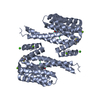
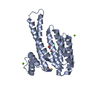
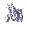
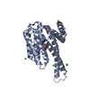
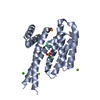
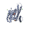
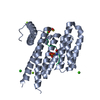
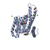
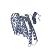
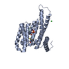
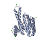
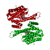
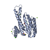
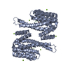
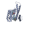
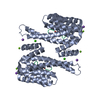
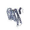
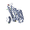
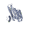
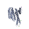
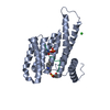
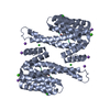
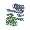
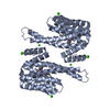
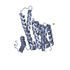
| タイトル | 14-3-3 Sigma in complex with phosphorylated (pS210) Gab2 peptide |
|---|
 要素 要素 | |
|---|
 キーワード キーワード | PROTEIN BINDING / 14-3-3 / Gab2 / complex / protein / protein-protein interactions |
|---|
| 機能・相同性 |  機能・相同性情報 機能・相同性情報
positive regulation of mast cell degranulation / STAT5 Activation / transmembrane receptor protein tyrosine kinase adaptor activity / phosphatidylinositol-3,4-bisphosphate binding / Signaling by cytosolic FGFR1 fusion mutants / Interleukin-15 signaling / STAT5 activation downstream of FLT3 ITD mutants / regulation of epidermal cell division / protein kinase C inhibitor activity / positive regulation of epidermal cell differentiation ...positive regulation of mast cell degranulation / STAT5 Activation / transmembrane receptor protein tyrosine kinase adaptor activity / phosphatidylinositol-3,4-bisphosphate binding / Signaling by cytosolic FGFR1 fusion mutants / Interleukin-15 signaling / STAT5 activation downstream of FLT3 ITD mutants / regulation of epidermal cell division / protein kinase C inhibitor activity / positive regulation of epidermal cell differentiation / keratinocyte development / keratinization / regulation of cell-cell adhesion / phosphatidylinositol-3,4,5-trisphosphate binding / RET signaling / cAMP/PKA signal transduction / Regulation of localization of FOXO transcription factors / PI3K Cascade / keratinocyte proliferation / phosphoserine residue binding / Role of LAT2/NTAL/LAB on calcium mobilization / Activation of BAD and translocation to mitochondria / Interleukin receptor SHC signaling / negative regulation of keratinocyte proliferation / establishment of skin barrier / negative regulation of protein localization to plasma membrane / Chk1/Chk2(Cds1) mediated inactivation of Cyclin B:Cdk1 complex / SARS-CoV-2 targets host intracellular signalling and regulatory pathways / negative regulation of protein kinase activity / negative regulation of stem cell proliferation / Signaling by CSF3 (G-CSF) / SARS-CoV-1 targets host intracellular signalling and regulatory pathways / RHO GTPases activate PKNs / Signaling by FLT3 ITD and TKD mutants / positive regulation of protein localization / Signaling by FLT3 fusion proteins / FLT3 Signaling / positive regulation of cell adhesion / protein sequestering activity / negative regulation of innate immune response / protein export from nucleus / osteoclast differentiation / TP53 Regulates Transcription of Genes Involved in G2 Cell Cycle Arrest / release of cytochrome c from mitochondria / positive regulation of protein export from nucleus / stem cell proliferation / Translocation of SLC2A4 (GLUT4) to the plasma membrane / TP53 Regulates Metabolic Genes / phosphatidylinositol 3-kinase/protein kinase B signal transduction / Signaling by SCF-KIT / Signaling by CSF1 (M-CSF) in myeloid cells / intrinsic apoptotic signaling pathway in response to DNA damage / Constitutive Signaling by Aberrant PI3K in Cancer / intracellular protein localization / PIP3 activates AKT signaling / regulation of protein localization / PI5P, PP2A and IER3 Regulate PI3K/AKT Signaling / positive regulation of cell growth / regulation of cell cycle / cadherin binding / membrane raft / positive regulation of cell population proliferation / protein kinase binding / negative regulation of transcription by RNA polymerase II / signal transduction / extracellular space / extracellular exosome / identical protein binding / nucleus / plasma membrane / cytosol / cytoplasm類似検索 - 分子機能 GRB2-associated-binding protein 1-4-like / 14-3-3 protein sigma / 14-3-3 proteins signature 2. / 14-3-3 protein, conserved site / 14-3-3 proteins signature 1. / 14-3-3 protein / 14-3-3 homologues / 14-3-3 domain / 14-3-3 domain superfamily / 14-3-3 protein ...GRB2-associated-binding protein 1-4-like / 14-3-3 protein sigma / 14-3-3 proteins signature 2. / 14-3-3 protein, conserved site / 14-3-3 proteins signature 1. / 14-3-3 protein / 14-3-3 homologues / 14-3-3 domain / 14-3-3 domain superfamily / 14-3-3 protein / PH domain / PH domain profile. / Pleckstrin homology domain. / Pleckstrin homology domain / PH-like domain superfamily類似検索 - ドメイン・相同性 14-3-3 protein sigma / GRB2-associated-binding protein 2類似検索 - 構成要素 |
|---|
| 生物種 |  Homo sapiens (ヒト) Homo sapiens (ヒト) |
|---|
| 手法 |  X線回折 / シンクロトロン / 分子置換 / 解像度: 1.95 Å X線回折 / シンクロトロン / 分子置換 / 解像度: 1.95 Å |
|---|
 データ登録者 データ登録者 | Ballone, A. / Lau, R.A. / Zweipfenning, F.P.A. / Ottmann, C. |
|---|
| 資金援助 |  オランダ, 1件 オランダ, 1件 | 組織 | 認可番号 | 国 |
|---|
| Marie Sklodowska-Curie Actions, FragNET ITN | 675179 | オランダ |
|
|---|
 引用 引用 | ジャーナル: Acta Crystallogr.,Sect.F / 年: 2020
タイトル: A new soaking procedure for X-ray crystallographic structural determination of protein-peptide complexes.
著者: Ballone, A. / Lau, R.A. / Zweipfenning, F.P.A. / Ottmann, C. |
|---|
| 履歴 | | 登録 | 2020年2月18日 | 登録サイト: PDBE / 処理サイト: PDBE |
|---|
| 改定 1.0 | 2020年10月14日 | Provider: repository / タイプ: Initial release |
|---|
| 改定 1.1 | 2024年1月24日 | Group: Advisory / Data collection ...Advisory / Data collection / Database references / Refinement description
カテゴリ: chem_comp_atom / chem_comp_bond ...chem_comp_atom / chem_comp_bond / database_2 / pdbx_initial_refinement_model / pdbx_unobs_or_zero_occ_residues
Item: _database_2.pdbx_DOI / _database_2.pdbx_database_accession |
|---|
| 改定 1.2 | 2024年11月20日 | Group: Structure summary
カテゴリ: pdbx_entry_details / pdbx_modification_feature
Item: _pdbx_entry_details.has_protein_modification |
|---|
|
|---|
 ムービー
ムービー コントローラー


 データを開く
 基本情報
基本情報 構造の表示
構造の表示 ダウンロードとリンク
 その他のダウンロード


 PDBj
PDBj


 集合体
集合体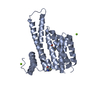


 分子量: 24.305 Da / 分子数: 2 / 由来タイプ: 合成 / 式: Mg
 分子量: 18.015 Da / 分子数: 432 / 由来タイプ: 天然 / 式: H2O
 試料調製
試料調製 / ビームライン: I03 / 波長: 0.973 Å
 解析
解析