 Movie
Movie Controller

[English] 日本語
 Yorodumi
- PDB-6xy9: Crystal structure of haloalkane dehalogenase DbeA-M1 loop variant... -
+ Open data

- Basic information
| Entry | Database: PDB / ID: 6xy9 | |||||||||||||||
|---|---|---|---|---|---|---|---|---|---|---|---|---|---|---|---|---|
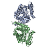
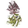
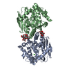
| Title | Crystal structure of haloalkane dehalogenase DbeA-M1 loop variant from Bradyrhizobium elkanii | |||||||||||||||
 Components Components | Haloalkane dehalogenase | |||||||||||||||
 Keywords Keywords | HYDROLASE / Haloalakane dehalogenase / microbial hydrolase / loop-helix engineering | |||||||||||||||
| Function / homology |  Function and homology information Function and homology information | |||||||||||||||
| Biological species |  Bradyrhizobium elkanii USDA 94 (bacteria) Bradyrhizobium elkanii USDA 94 (bacteria) | |||||||||||||||
| Method |  X-RAY DIFFRACTION / SYNCHROTRON / MOLECULAR REPLACEMENT / Resolution: 2.2 Å X-RAY DIFFRACTION / SYNCHROTRON / MOLECULAR REPLACEMENT / Resolution: 2.2 Å | |||||||||||||||
 Authors Authors | Prudnikova, T. / Rezacova, P. / Kuta Smatanova, I. / Chaloupkova, R. / Damborsky, J. | |||||||||||||||
| Funding support |  Czech Republic, 4items Czech Republic, 4items
| |||||||||||||||
 Citation Citation | Journal: Comput Struct Biotechnol J / Year: 2020 Title: Structural and catalytic effects of surface loop-helix transplantation within haloalkane dehalogenase family. Authors: Marek, M. / Chaloupkova, R. / Prudnikova, T. / Sato, Y. / Rezacova, P. / Nagata, Y. / Kuta Smatanova, I. / Damborsky, J. | |||||||||||||||
| History |
|
- Structure visualization
| Structure viewer | Molecule: MolmilJmol/JSmol |
|---|
- Downloads & links
-Download
| PDBx/mmCIF format | 6xy9.cif.gz | 135.9 KB | Display | PDBx/mmCIF format |
|---|---|---|---|---|
| PDB format | pdb6xy9.ent.gz | 104.7 KB | Display | PDB format |
| PDBx/mmJSON format | 6xy9.json.gz | Tree view | PDBx/mmJSON format | |
| Others |  Other downloads Other downloads |
-Validation report
| Arichive directory | https://data.pdbj.org/pub/pdb/validation_reports/xy/6xy9ftp://data.pdbj.org/pub/pdb/validation_reports/xy/6xy9 | HTTPS FTP |
|---|
-Related structure data
| Related structure data | 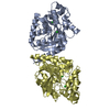 4k2aS S: Starting model for refinement |
|---|---|
| Similar structure data |


-Links
 PDBj
PDBj


- Assembly
| Deposited unit | 
| ||||||||
|---|---|---|---|---|---|---|---|---|---|
| 1 |
| ||||||||
| Unit cell |
| ||||||||
| Components on special symmetry positions |
|
-Components
| #1: Protein | Mass: 33731.242 Da / Num. of mol.: 2 / Mutation: 143VAEEQDHAE Source method: isolated from a genetically manipulated source Details: artifitially synthesized Source: (gene. exp.) Bradyrhizobium elkanii USDA 94 (bacteria)Gene: dbeA, dhaA / Plasmid: pET-21b / Production host: #2: Chemical | ChemComp-CL /   Mass: 35.453 Da / Num. of mol.: 4 / Source method: obtained synthetically / Formula: Cl Mass: 35.453 Da / Num. of mol.: 4 / Source method: obtained synthetically / Formula: Cl#3: Chemical |   Mass: 59.044 Da / Num. of mol.: 2 / Source method: obtained synthetically / Formula: C2H3O2 / Feature type: SUBJECT OF INVESTIGATION Mass: 59.044 Da / Num. of mol.: 2 / Source method: obtained synthetically / Formula: C2H3O2 / Feature type: SUBJECT OF INVESTIGATION#4: Water | ChemComp-HOH / |  Mass: 18.015 Da / Num. of mol.: 352 / Source method: isolated from a natural source / Formula: H2O Mass: 18.015 Da / Num. of mol.: 352 / Source method: isolated from a natural source / Formula: H2OHas ligand of interest | Y | |
|---|
-Experimental details
-Experiment
| Experiment | Method: X-RAY DIFFRACTION / Number of used crystals: 1 |
|---|
- Sample preparation
| Crystal | Density Matthews: 2.89 Å3/Da / Density % sol: 45 % |
|---|---|
| Crystal grow | Temperature: 277 K / Method: vapor diffusion, sitting drop / pH: 7.5 / Details: 25% PEG 4000, 130 mM calcium acetate |
-Data collection
| Diffraction | Mean temperature: 100 K / Serial crystal experiment: N |
|---|---|
| Diffraction source | Source: SYNCHROTRON / Site: EMBL/DESY, HAMBURG  / Beamline: X11 / Wavelength: 0.918 Å / Beamline: X11 / Wavelength: 0.918 Å |
| Detector | Type: MAR555 FLAT PANEL / Detector: IMAGE PLATE / Date: Jan 16, 2008 |
| Radiation | Protocol: SINGLE WAVELENGTH / Monochromatic (M) / Laue (L): M / Scattering type: x-ray |
| Radiation wavelength | Wavelength: 0.918 Å / Relative weight: 1 |
| Reflection | Resolution: 2.2→19.6 Å / Num. obs: 38810 / % possible obs: 98.6 % / Redundancy: 3.74 % / Biso Wilson estimate: 32.3 Å2 / CC1/2: 0.86 / Rmerge(I) obs: 0.138 / Net I/σ(I): 11.76 |
| Reflection shell | Resolution: 2.2→2.27 Å / Rmerge(I) obs: 0.515 / Mean I/σ(I) obs: 3.35 / Num. unique obs: 6037 / CC1/2: 0.628 |
- Processing
| Software |
| |||||||||||||||||||||||||||||||||||||||||||||
|---|---|---|---|---|---|---|---|---|---|---|---|---|---|---|---|---|---|---|---|---|---|---|---|---|---|---|---|---|---|---|---|---|---|---|---|---|---|---|---|---|---|---|---|---|---|---|
| Refinement | Method to determine structure: MOLECULAR REPLACEMENT Starting model: 4k2a Resolution: 2.2→19.6 Å / Cor.coef. Fo:Fc: 0.949 / Cor.coef. Fo:Fc free: 0.902 / SU B: 5.459 / SU ML: 0.136 / Cross valid method: FREE R-VALUE / σ(F): 0 / ESU R: 0.216 / ESU R Free: 0.196 / Details: U VALUES : REFINED INDIVIDUALLY
| |||||||||||||||||||||||||||||||||||||||||||||
| Solvent computation | Ion probe radii: 0.8 Å / Shrinkage radii: 0.8 Å / VDW probe radii: 1.2 Å | |||||||||||||||||||||||||||||||||||||||||||||
| Displacement parameters | Biso max: 124.2 Å2 / Biso mean: 31.675 Å2 / Biso min: 12.16 Å2
| |||||||||||||||||||||||||||||||||||||||||||||
| Refinement step | Cycle: final / Resolution: 2.2→19.6 Å
| |||||||||||||||||||||||||||||||||||||||||||||
| Refine LS restraints |
| |||||||||||||||||||||||||||||||||||||||||||||
| LS refinement shell | Resolution: 2.2→2.256 Å / Rfactor Rfree error: 0 / Total num. of bins used: 20
|
