 Movie
Movie Controller


+ Open data

- Basic information
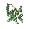
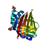
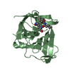
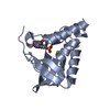
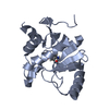
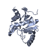
| Entry | Database: PDB / ID: 6xu5 | ||||||
|---|---|---|---|---|---|---|---|
| Title | Human myelin protein P2 mutant N2D | ||||||
 Components Components | Myelin P2 protein | ||||||
 Keywords Keywords | LIPID BINDING PROTEIN / mutant / peripheral membrane protein / FABP / beta barrel | ||||||
| Function / homology |  Function and homology information Function and homology informationmembrane organization / cholesterol binding / fatty acid transport / fatty acid binding / myelin sheath / extracellular exosome / nucleus / cytosol Similarity search - Function | ||||||
| Biological species |  Homo sapiens (human) Homo sapiens (human) | ||||||
| Method |  X-RAY DIFFRACTION / SYNCHROTRON / MOLECULAR REPLACEMENT / Resolution: 1.65 Å X-RAY DIFFRACTION / SYNCHROTRON / MOLECULAR REPLACEMENT / Resolution: 1.65 Å | ||||||
 Authors Authors | Ruskamo, S. / Lehtimaki, M. / Kursula, P. | ||||||
| Funding support |  Finland, 1items Finland, 1items
| ||||||
 Citation Citation | Journal: J.Biol.Chem. / Year: 2020 Title: Cryo-EM, X-ray diffraction, and atomistic simulations reveal determinants for the formation of a supramolecular myelin-like proteolipid lattice. Authors: Ruskamo, S. / Krokengen, O.C. / Kowal, J. / Nieminen, T. / Lehtimaki, M. / Raasakka, A. / Dandey, V.P. / Vattulainen, I. / Stahlberg, H. / Kursula, P. | ||||||
| History |
|
- Structure visualization
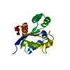
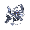
| Structure viewer | Molecule: MolmilJmol/JSmol |
|---|
- Downloads & links
-Download
| PDBx/mmCIF format | 6xu5.cif.gz | 79.9 KB | Display | PDBx/mmCIF format |
|---|---|---|---|---|
| PDB format | pdb6xu5.ent.gz | 59.3 KB | Display | PDB format |
| PDBx/mmJSON format | 6xu5.json.gz | Tree view | PDBx/mmJSON format | |
| Others |  Other downloads Other downloads |
-Validation report
| Arichive directory | https://data.pdbj.org/pub/pdb/validation_reports/xu/6xu5ftp://data.pdbj.org/pub/pdb/validation_reports/xu/6xu5 | HTTPS FTP |
|---|
-Related structure data
| Related structure data |  6stsC  6xu9C  6xuaC 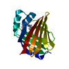 6xuwC  6xvqC  6xvrC  6xvsC  6xvyC  6xw9C  2wutS S: Starting model for refinement C: citing same article ( |
|---|---|
| Similar structure data |


-Links
 PDBj
PDBj


- Assembly
| Deposited unit | 
| ||||||||||||
|---|---|---|---|---|---|---|---|---|---|---|---|---|---|
| 1 |
| ||||||||||||
| Unit cell |
|
-Components
| #1: Protein | Mass: 14992.456 Da / Num. of mol.: 1 / Mutation: N2D Source method: isolated from a genetically manipulated source Source: (gene. exp.) Homo sapiens (human) / Gene: PMP2 / Production host:  | ||||
|---|---|---|---|---|---|
| #2: Chemical | ChemComp-PLM /   Mass: 256.424 Da / Num. of mol.: 1 / Source method: obtained synthetically / Formula: C16H32O2 Mass: 256.424 Da / Num. of mol.: 1 / Source method: obtained synthetically / Formula: C16H32O2 | ||||
| #3: Chemical |   Mass: 192.124 Da / Num. of mol.: 2 / Source method: obtained synthetically / Formula: C6H8O7 Mass: 192.124 Da / Num. of mol.: 2 / Source method: obtained synthetically / Formula: C6H8O7#4: Water | ChemComp-HOH / |  Mass: 18.015 Da / Num. of mol.: 286 / Source method: isolated from a natural source / Formula: H2O Mass: 18.015 Da / Num. of mol.: 286 / Source method: isolated from a natural source / Formula: H2OHas ligand of interest | N | |
-Experimental details
-Experiment
| Experiment | Method: X-RAY DIFFRACTION / Number of used crystals: 1 |
|---|
- Sample preparation
| Crystal | Density Matthews: 2.88 Å3/Da / Density % sol: 57.27 % |
|---|---|
| Crystal grow | Temperature: 277 K / Method: vapor diffusion, sitting drop / pH: 5.5 / Details: 36% PEG 6000, 0.1 M citrate pH 5.5 |
-Data collection
| Diffraction | Mean temperature: 100 K / Serial crystal experiment: N |
|---|---|
| Diffraction source | Source: SYNCHROTRON / Site: EMBL/DESY, HAMBURG  / Beamline: X12 / Wavelength: 0.97 Å / Beamline: X12 / Wavelength: 0.97 Å |
| Detector | Type: RAYONIX MX225-HS / Detector: CCD / Date: Nov 19, 2010 |
| Radiation | Protocol: SINGLE WAVELENGTH / Monochromatic (M) / Laue (L): M / Scattering type: x-ray |
| Radiation wavelength | Wavelength: 0.97 Å / Relative weight: 1 |
| Reflection | Resolution: 1.65→30 Å / Num. obs: 21068 / % possible obs: 97.4 % / Redundancy: 4.7 % / Biso Wilson estimate: 19 Å2 / CC1/2: 0.998 / Rrim(I) all: 0.09 / Rsym value: 0.08 / Net I/σ(I): 15.2 |
| Reflection shell | Resolution: 1.65→1.7 Å / Redundancy: 2 % / Mean I/σ(I) obs: 1.5 / Num. unique obs: 2445 / CC1/2: 0.625 / Rrim(I) all: 0.703 / Rsym value: 0.541 / % possible all: 78.9 |
- Processing
| Software |
| ||||||||||||||||||||||||
|---|---|---|---|---|---|---|---|---|---|---|---|---|---|---|---|---|---|---|---|---|---|---|---|---|---|
| Refinement | Method to determine structure: MOLECULAR REPLACEMENT Starting model: 2wut Resolution: 1.65→30 Å / Cross valid method: FREE R-VALUE
| ||||||||||||||||||||||||
| Displacement parameters | Biso mean: 15.15 Å2 | ||||||||||||||||||||||||
| Refinement step | Cycle: LAST / Resolution: 1.65→30 Å
| ||||||||||||||||||||||||
| Refine LS restraints |
|
