National Institutes of Health/National Institute on Aging (NIH/NIA)
R01 GM121964
米国
引用
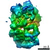
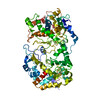
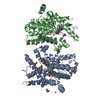
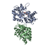
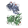
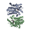
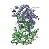
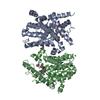
ジャーナル: Structure / 年: 2021 タイトル: Structural analysis of Mycobacterium tuberculosis M13 metalloprotease Zmp1 open states. 著者: Wenguang G Liang / Jordan M Mancl / Minglei Zhao / Wei-Jen Tang / 要旨: Zinc metalloprotease 1 (Zmp1), a Mycobacterium tuberculosis 75 kDa secreted enzyme, mediates key stages of tuberculosis disease progression. The biological activity of Zmp1 presumably stems from its ...Zinc metalloprotease 1 (Zmp1), a Mycobacterium tuberculosis 75 kDa secreted enzyme, mediates key stages of tuberculosis disease progression. The biological activity of Zmp1 presumably stems from its ability to degrade bacterium- and/or host-derived peptides. The crystal structures of Zmp1 and related M13 metalloproteases, such as neprilysin and endothelin-converting enzyme-1 were determined only in the closed conformation, which cannot capture substrates or release proteolytic products. Thus, the mechanisms of substrate binding and selectivity remain elusive. Here we report two open-state cryo-EM structures of Zmp1, revealed by our SAXS analysis to be the dominant states in solution. Our structural analyses reveal how ligand binding induces a conformational switch in four linker regions to drive the rigid body motion of the D1 and D2 domains, which form the sizable catalytic chamber. Furthermore, they offer insights into the catalytic cycle and mechanism of substrate recognition of M13 metalloproteases for future therapeutic innovations.
履歴
登録
2020年6月29日
登録サイト: RCSB / 処理サイト: RCSB
改定 1.0
2020年12月23日
Provider: repository / タイプ: Initial release
改定 1.0
2020年12月23日
Data content type: EM metadata / Data content type: EM metadata / Provider: repository / タイプ: Initial release
改定 1.0
2020年12月23日
Data content type: Additional map / Data content type: Additional map / Provider: repository / タイプ: Initial release
改定 1.0
2020年12月23日
Data content type: FSC / Data content type: FSC / Provider: repository / タイプ: Initial release
改定 1.0
2020年12月23日
Data content type: Half map / Part number: 1 / Data content type: Half map / Provider: repository / タイプ: Initial release
改定 1.0
2020年12月23日
Data content type: Half map / Part number: 2 / Data content type: Half map / Provider: repository / タイプ: Initial release
改定 1.0
2020年12月23日
Data content type: Image / Data content type: Image / Provider: repository / タイプ: Initial release
改定 1.0
2020年12月23日
Data content type: Mask / Data content type: Mask / Provider: repository / タイプ: Initial release
改定 1.0
2020年12月23日
Data content type: Primary map / Data content type: Primary map / Provider: repository / タイプ: Initial release
改定 1.0
2020年12月23日
Data content type: Additional map / Data content type: Additional map / Provider: repository / タイプ: Initial release
改定 1.0
2020年12月23日
Data content type: FSC / Data content type: FSC / Provider: repository / タイプ: Initial release
改定 1.0
2020年12月23日
Data content type: Half map / Part number: 1 / Data content type: Half map / Provider: repository / タイプ: Initial release
改定 1.0
2020年12月23日
Data content type: Half map / Part number: 2 / Data content type: Half map / Provider: repository / タイプ: Initial release
改定 1.0
2020年12月23日
Data content type: Image / Data content type: Image / Provider: repository / タイプ: Initial release
改定 1.0
2020年12月23日
Data content type: Mask / Data content type: Mask / Provider: repository / タイプ: Initial release
改定 1.0
2020年12月23日
Data content type: Primary map / Data content type: Primary map / Provider: repository / タイプ: Initial release
Data content type: EM metadata / Data content type: EM metadata / EM metadata / Group: Data processing / Experimental summary / Data content type: EM metadata / EM metadata / カテゴリ: em_admin / em_software / Data content type: EM metadata / EM metadata / Item: _em_admin.last_update / _em_software.name
 ムービー
ムービー コントローラー


 データを開く
 基本情報
基本情報 要素
要素 キーワード
キーワード 機能・相同性情報
 Mycobacterium tuberculosis (結核菌)
 データ登録者
 米国, 1件
  引用
引用 構造の表示
構造の表示 ダウンロードとリンク
 その他のダウンロード
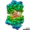


 PDBj
PDBj


 集合体
集合体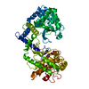

 分子量: 65.409 Da / 分子数: 1 / 由来タイプ: 合成 / 式: Zn
 試料調製
試料調製 電子顕微鏡撮影
 FIELD EMISSION GUN / 加速電圧: 300 kV / 照射モード: FLOOD BEAM
 解析
解析