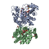
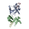
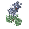
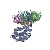
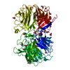
Entry Database : PDB / ID : 6fqgTitle GluA2(flop) G724C ligand binding core dimer bound to L-Glutamate (Form A) at 2.34 Angstrom resolution Glutamate receptor 2 Keywords / / / / Function / homology Function Domain/homology Component
/ / / / / / / / / / / / / / / / / / / / / / / / / / / / / / / / / / / / / / / / / / / / / / / / / / / / / / / / / / / / / / / / / / / / / / / / / / / / / / / / / / / / / / / / / / / / / / / / / / / / / / / / / / / / / / / / / / / Biological species Rattus norvegicus (Norway rat)Method / / / / Resolution : 2.34139094861 Å Authors Coombs, I.D. / Soto, D. / Gold, M.G. / Farrant, M.F. / Cull-Candy, S.G. Funding support Organization Grant number Country Medical Research Council (United Kingdom) MR/J002976/1 Medical Research Council (United Kingdom) MR/J012998/1 Wellcome Trust 086185/Z/08/Z
Journal : To Be Published Title : X-ray structure of GluA2 flop G724C ligand binding core dimer bound to glutamate at 2.32 Angstroms resolutionAuthors : Coombs, I.D. / Soto, D. / Gold, M.G. / Farrant, M.F. / Cull-Candy, S.G. History Deposition Feb 14, 2018 Deposition site / Processing site Revision 1.0 Mar 13, 2019 Provider / Type Revision 2.0 Mar 15, 2023 Group Atomic model / Data collection ... Atomic model / Data collection / Database references / Derived calculations / Non-polymer description / Source and taxonomy / Structure summary Category atom_site / chem_comp ... atom_site / chem_comp / database_2 / entity / entity_name_com / entity_src_gen / pdbx_entity_nonpoly / pdbx_nonpoly_scheme / struct_site Item _atom_site.auth_comp_id / _atom_site.label_comp_id ... _atom_site.auth_comp_id / _atom_site.label_comp_id / _chem_comp.formula / _chem_comp.formula_weight / _chem_comp.id / _chem_comp.mon_nstd_flag / _chem_comp.name / _chem_comp.pdbx_synonyms / _chem_comp.type / _database_2.pdbx_DOI / _database_2.pdbx_database_accession / _entity.pdbx_description / _entity.pdbx_mutation / _entity_name_com.name / _entity_src_gen.gene_src_common_name / _pdbx_entity_nonpoly.comp_id / _pdbx_entity_nonpoly.name / _pdbx_nonpoly_scheme.mon_id / _pdbx_nonpoly_scheme.pdb_mon_id / _struct_site.details / _struct_site.pdbx_auth_comp_id Revision 2.1 Feb 7, 2024 Group / Refinement descriptionCategory chem_comp_atom / chem_comp_bond ... chem_comp_atom / chem_comp_bond / pdbx_initial_refinement_model / struct_ncs_dom_lim Item _struct_ncs_dom_lim.beg_auth_comp_id / _struct_ncs_dom_lim.beg_label_asym_id ... _struct_ncs_dom_lim.beg_auth_comp_id / _struct_ncs_dom_lim.beg_label_asym_id / _struct_ncs_dom_lim.beg_label_comp_id / _struct_ncs_dom_lim.beg_label_seq_id / _struct_ncs_dom_lim.end_auth_comp_id / _struct_ncs_dom_lim.end_label_asym_id / _struct_ncs_dom_lim.end_label_comp_id / _struct_ncs_dom_lim.end_label_seq_id Revision 2.2 Oct 23, 2024 Group / Category / pdbx_modification_feature
Show all Show less
 Movie
Movie Controller

 Yorodumi
 Open data
 Basic information
 Components
 Keywords
 Function and homology information
 X-RAY DIFFRACTION /
  Authors
 United Kingdom, 3items
  Citation
 Structure visualization
 Downloads & links
 Other downloads
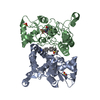


 PDBj
PDBj


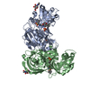
 Assembly
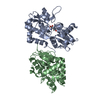


 Type: L-peptide linking / Mass: 147.129 Da / Num. of mol.: 2 / Source method: obtained synthetically / Formula: C5H9NO4
 Mass: 18.015 Da / Num. of mol.: 104 / Source method: isolated from a natural source / Formula: H2O
 Sample preparation
 Processing