 Movie
Movie Controller

[English] 日本語
 Yorodumi
- PDB-6xh5: Hierarchical design of multi-scale protein complexes by combinato... -
+ Open data

- Basic information
| Entry | Database: PDB / ID: 6xh5 | ||||||
|---|---|---|---|---|---|---|---|
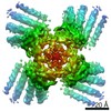
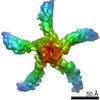
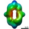
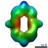
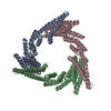
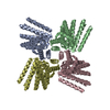
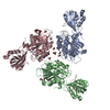
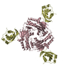
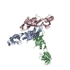
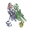
| Title | Hierarchical design of multi-scale protein complexes by combinatorial assembly of oligomeric helical bundle and repeat protein building blocks | ||||||
 Components Components | helical fusion design | ||||||
 Keywords Keywords | DE NOVO PROTEIN / helical fusion / de novo design | ||||||
| Function / homology | Aldolase class I / TIM Barrel / Alpha-Beta Barrel / Alpha Beta Function and homology information Function and homology information | ||||||
| Biological species | synthetic construct (others) | ||||||
| Method |  X-RAY DIFFRACTION / SYNCHROTRON / MOLECULAR REPLACEMENT / Resolution: 3.32 Å X-RAY DIFFRACTION / SYNCHROTRON / MOLECULAR REPLACEMENT / Resolution: 3.32 Å | ||||||
 Authors Authors | Bera, A.K. / Hsia, Y. / Kang, A.S. / Baker, D. | ||||||
| Funding support |  United States, 1items United States, 1items
| ||||||
 Citation Citation | Journal: Nat Commun / Year: 2021 Title: Design of multi-scale protein complexes by hierarchical building block fusion. Authors: Yang Hsia / Rubul Mout / William Sheffler / Natasha I Edman / Ivan Vulovic / Young-Jun Park / Rachel L Redler / Matthew J Bick / Asim K Bera / Alexis Courbet / Alex Kang / T J Brunette / Una ...Authors: Yang Hsia / Rubul Mout / William Sheffler / Natasha I Edman / Ivan Vulovic / Young-Jun Park / Rachel L Redler / Matthew J Bick / Asim K Bera / Alexis Courbet / Alex Kang / T J Brunette / Una Nattermann / Evelyn Tsai / Ayesha Saleem / Cameron M Chow / Damian Ekiert / Gira Bhabha / David Veesler / David Baker / Abstract: A systematic and robust approach to generating complex protein nanomaterials would have broad utility. We develop a hierarchical approach to designing multi-component protein assemblies from two ...A systematic and robust approach to generating complex protein nanomaterials would have broad utility. We develop a hierarchical approach to designing multi-component protein assemblies from two classes of modular building blocks: designed helical repeat proteins (DHRs) and helical bundle oligomers (HBs). We first rigidly fuse DHRs to HBs to generate a large library of oligomeric building blocks. We then generate assemblies with cyclic, dihedral, and point group symmetries from these building blocks using architecture guided rigid helical fusion with new software named WORMS. X-ray crystallography and cryo-electron microscopy characterization show that the hierarchical design approach can accurately generate a wide range of assemblies, including a 43 nm diameter icosahedral nanocage. The computational methods and building block sets described here provide a very general route to de novo designed protein nanomaterials. | ||||||
| History |
|
- Structure visualization
| Structure viewer | Molecule: MolmilJmol/JSmol |
|---|
- Downloads & links
-Download
| PDBx/mmCIF format | 6xh5.cif.gz | 954.2 KB | Display | PDBx/mmCIF format |
|---|---|---|---|---|
| PDB format | pdb6xh5.ent.gz | 674.7 KB | Display | PDB format |
| PDBx/mmJSON format | 6xh5.json.gz | Tree view | PDBx/mmJSON format | |
| Others |  Other downloads Other downloads |
-Validation report
| Arichive directory | https://data.pdbj.org/pub/pdb/validation_reports/xh/6xh5ftp://data.pdbj.org/pub/pdb/validation_reports/xh/6xh5 | HTTPS FTP |
|---|
-Related structure data


-Links
 PDBj
PDBj
- Assembly
| Deposited unit | 
| ||||||||||||
|---|---|---|---|---|---|---|---|---|---|---|---|---|---|
| 1 | 
| ||||||||||||
| 2 | 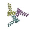
| ||||||||||||
| Unit cell |
|
-Components
| #1: Protein | Mass: 39119.957 Da / Num. of mol.: 6 Source method: isolated from a genetically manipulated source Source: (gene. exp.) synthetic construct (others) / Production host:  #2: Water | ChemComp-HOH / |  Mass: 18.015 Da / Num. of mol.: 46 / Source method: isolated from a natural source / Formula: H2O Mass: 18.015 Da / Num. of mol.: 46 / Source method: isolated from a natural source / Formula: H2O |
|---|
-Experimental details
-Experiment
| Experiment | Method: X-RAY DIFFRACTION / Number of used crystals: 1 |
|---|
- Sample preparation
| Crystal | Density Matthews: 3.31 Å3/Da / Density % sol: 62.84 % |
|---|---|
| Crystal grow | Temperature: 293 K / Method: vapor diffusion, sitting drop Details: 0.2 M Lithium sulfate, 0.1 Na-acetate pH 4.5 and 2.5 M NaCl |
-Data collection
| Diffraction | Mean temperature: 100 K / Serial crystal experiment: N |
|---|---|
| Diffraction source | Source: SYNCHROTRON / Site: APS / Beamline: 24-ID-E / Wavelength: 0.97918 Å |
| Detector | Type: DECTRIS PILATUS 6M / Detector: PIXEL / Date: May 29, 2020 |
| Radiation | Protocol: SINGLE WAVELENGTH / Monochromatic (M) / Laue (L): M / Scattering type: x-ray |
| Radiation wavelength | Wavelength: 0.97918 Å / Relative weight: 1 |
| Reflection | Resolution: 3.32→78.12 Å / Num. obs: 47181 / % possible obs: 99.86 % / Redundancy: 27.2 % / Biso Wilson estimate: 116.55 Å2 / CC1/2: 0.986 / Rpim(I) all: 0.0463 / Net I/σ(I): 17.98 |
| Reflection shell | Resolution: 3.32→3.43 Å / Mean I/σ(I) obs: 2.09 / Num. unique obs: 4621 / CC1/2: 0.723 / Rpim(I) all: 0.3488 / % possible all: 28.4 |
- Processing
| Software |
| ||||||||||||||||||||||||||||||||||||||||||||||||||||||||||||||||||||||||||||||||||||||||||||||||||||||||||||||||||||||||||||||
|---|---|---|---|---|---|---|---|---|---|---|---|---|---|---|---|---|---|---|---|---|---|---|---|---|---|---|---|---|---|---|---|---|---|---|---|---|---|---|---|---|---|---|---|---|---|---|---|---|---|---|---|---|---|---|---|---|---|---|---|---|---|---|---|---|---|---|---|---|---|---|---|---|---|---|---|---|---|---|---|---|---|---|---|---|---|---|---|---|---|---|---|---|---|---|---|---|---|---|---|---|---|---|---|---|---|---|---|---|---|---|---|---|---|---|---|---|---|---|---|---|---|---|---|---|---|---|---|
| Refinement | Method to determine structure: MOLECULAR REPLACEMENT Starting model: Designed model Resolution: 3.32→78.12 Å / SU ML: 0.4571 / Cross valid method: FREE R-VALUE / σ(F): 1.34 / Phase error: 28.3973 Stereochemistry target values: GeoStd + Monomer Library + CDL v1.2
| ||||||||||||||||||||||||||||||||||||||||||||||||||||||||||||||||||||||||||||||||||||||||||||||||||||||||||||||||||||||||||||||
| Solvent computation | Shrinkage radii: 0.9 Å / VDW probe radii: 1.11 Å / Solvent model: FLAT BULK SOLVENT MODEL | ||||||||||||||||||||||||||||||||||||||||||||||||||||||||||||||||||||||||||||||||||||||||||||||||||||||||||||||||||||||||||||||
| Displacement parameters | Biso mean: 117.39 Å2 | ||||||||||||||||||||||||||||||||||||||||||||||||||||||||||||||||||||||||||||||||||||||||||||||||||||||||||||||||||||||||||||||
| Refinement step | Cycle: LAST / Resolution: 3.32→78.12 Å
| ||||||||||||||||||||||||||||||||||||||||||||||||||||||||||||||||||||||||||||||||||||||||||||||||||||||||||||||||||||||||||||||
| Refine LS restraints |
| ||||||||||||||||||||||||||||||||||||||||||||||||||||||||||||||||||||||||||||||||||||||||||||||||||||||||||||||||||||||||||||||
| LS refinement shell |
| ||||||||||||||||||||||||||||||||||||||||||||||||||||||||||||||||||||||||||||||||||||||||||||||||||||||||||||||||||||||||||||||
| Refinement TLS params. | Method: refined / Origin x: -18.1497037018 Å / Origin y: -16.0583704885 Å / Origin z: 55.7190702377 Å
| ||||||||||||||||||||||||||||||||||||||||||||||||||||||||||||||||||||||||||||||||||||||||||||||||||||||||||||||||||||||||||||||
| Refinement TLS group | Selection details: all |
