 Movie
Movie Controller

[English] 日本語
 Yorodumi
- PDB-6hc7: The crystal structure of BSAP, a zinc aminopeptidase from Bacillu... -
+ Open data

- Basic information
| Entry | Database: PDB / ID: 6hc7 | ||||||
|---|---|---|---|---|---|---|---|
| Title | The crystal structure of BSAP, a zinc aminopeptidase from Bacillus subtilis (medium resolution) | ||||||
 Components Components | Aminopeptidase Y (Arg Lys Leu preference) | ||||||
 Keywords Keywords | METAL BINDING PROTEIN / aminopeptidase / bacillus subtilis / PA-domain / zinc enzyme | ||||||
| Function / homology |  Function and homology information Function and homology informationaminopeptidase B / bacterial leucyl aminopeptidase / metalloexopeptidase activity / aminopeptidase activity / proteolysis / extracellular region / metal ion binding Similarity search - Function | ||||||
| Biological species |  | ||||||
| Method |  X-RAY DIFFRACTION / SYNCHROTRON / MOLECULAR REPLACEMENT / Resolution: 2.5 Å X-RAY DIFFRACTION / SYNCHROTRON / MOLECULAR REPLACEMENT / Resolution: 2.5 Å | ||||||
 Authors Authors | Alhadeff, R. / Lansky, S. / Feinberg, H. / Shoham, Y. / Shoham, G. | ||||||
 Citation Citation | Journal: To Be Published Title: The crystal structure of BSAP, a zinc aminopeptidase from Bacillus subtilis (medium resolution) Authors: Alhadeff, R. / Faygenboim, R. / Lansky, S. / Rogoulenko, E. / Cohen, T. / Fundoiano-Hershcovitz, Y. / Feinberg, H. / Shoham, Y. / Shoham, G. | ||||||
| History |
|
- Structure visualization
| Structure viewer | Molecule: MolmilJmol/JSmol |
|---|
- Downloads & links
-Download
| PDBx/mmCIF format | 6hc7.cif.gz | 249.1 KB | Display | PDBx/mmCIF format |
|---|---|---|---|---|
| PDB format | pdb6hc7.ent.gz | 200.3 KB | Display | PDB format |
| PDBx/mmJSON format | 6hc7.json.gz | Tree view | PDBx/mmJSON format | |
| Others |  Other downloads Other downloads |
-Validation report
| Arichive directory | https://data.pdbj.org/pub/pdb/validation_reports/hc/6hc7ftp://data.pdbj.org/pub/pdb/validation_reports/hc/6hc7 | HTTPS FTP |
|---|
-Related structure data
| Related structure data | 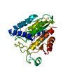 1cp7S S: Starting model for refinement |
|---|---|
| Similar structure data |


-Links
 PDBj
PDBj
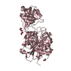
- Assembly
| Deposited unit | 
| ||||||||
|---|---|---|---|---|---|---|---|---|---|
| 1 | 
| ||||||||
| 2 | 
| ||||||||
| 3 | 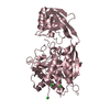
| ||||||||
| 4 |
| ||||||||
| Unit cell |
|
-Components
-Protein , 1 types, 3 molecules ABC
| #1: Protein | Mass: 49515.082 Da / Num. of mol.: 3 Source method: isolated from a genetically manipulated source Source: (gene. exp.) |
|---|
-Non-polymers , 5 types, 364 molecules 


| #2: Chemical | ChemComp-ZN /  Mass: 65.409 Da / Num. of mol.: 6 / Source method: obtained synthetically / Formula: Zn Mass: 65.409 Da / Num. of mol.: 6 / Source method: obtained synthetically / Formula: Zn#3: Chemical |  Mass: 96.063 Da / Num. of mol.: 3 / Source method: obtained synthetically / Formula: SO4 Mass: 96.063 Da / Num. of mol.: 3 / Source method: obtained synthetically / Formula: SO4#4: Chemical |  Mass: 59.044 Da / Num. of mol.: 3 / Source method: obtained synthetically / Formula: C2H3O2 Mass: 59.044 Da / Num. of mol.: 3 / Source method: obtained synthetically / Formula: C2H3O2#5: Chemical | ChemComp-CL /  Mass: 35.453 Da / Num. of mol.: 6 / Source method: obtained synthetically / Formula: Cl Mass: 35.453 Da / Num. of mol.: 6 / Source method: obtained synthetically / Formula: Cl#6: Water | ChemComp-HOH / | Mass: 18.015 Da / Num. of mol.: 346 / Source method: isolated from a natural source / Formula: H2O |
|---|
-Experimental details
-Experiment
| Experiment | Method: X-RAY DIFFRACTION / Number of used crystals: 1 |
|---|
- Sample preparation
| Crystal | Density Matthews: 2.11 Å3/Da / Density % sol: 41.81 % |
|---|---|
| Crystal grow | Temperature: 293 K / Method: vapor diffusion, hanging drop / pH: 5.6 Details: 20% PEG 2K MME, 0.1M LiSO4, 4mM MnCl2, 0.1M acetate pH 5.6 |
-Data collection
| Diffraction | Mean temperature: 100 K |
|---|---|
| Diffraction source | Source: SYNCHROTRON / Site: ESRF  / Beamline: ID14-4 / Wavelength: 1.282 Å / Beamline: ID14-4 / Wavelength: 1.282 Å |
| Detector | Type: ADSC QUANTUM 4 / Detector: CCD / Date: Jul 15, 2002 |
| Radiation | Protocol: SINGLE WAVELENGTH / Monochromatic (M) / Laue (L): M / Scattering type: x-ray |
| Radiation wavelength | Wavelength: 1.282 Å / Relative weight: 1 |
| Reflection | Resolution: 2.5→74 Å / Num. obs: 43610 / % possible obs: 98.8 % / Redundancy: 3.4 % / Rmerge(I) obs: 0.053 / Net I/σ(I): 13.1 |
| Reflection shell | Resolution: 2.5→2.54 Å / Rmerge(I) obs: 0.25 / % possible all: 86.3 |
- Processing
| Software |
| ||||||||||||||||||||||||||||||||||||||||||||||||||||||||||||||||||||||||||||||||||||||||||||||||||||||||||||||||
|---|---|---|---|---|---|---|---|---|---|---|---|---|---|---|---|---|---|---|---|---|---|---|---|---|---|---|---|---|---|---|---|---|---|---|---|---|---|---|---|---|---|---|---|---|---|---|---|---|---|---|---|---|---|---|---|---|---|---|---|---|---|---|---|---|---|---|---|---|---|---|---|---|---|---|---|---|---|---|---|---|---|---|---|---|---|---|---|---|---|---|---|---|---|---|---|---|---|---|---|---|---|---|---|---|---|---|---|---|---|---|---|---|---|
| Refinement | Method to determine structure: MOLECULAR REPLACEMENT Starting model: 1CP7 Resolution: 2.5→73.921 Å / SU ML: 0.35 / Cross valid method: FREE R-VALUE / σ(F): 1.36 / Phase error: 26.15
| ||||||||||||||||||||||||||||||||||||||||||||||||||||||||||||||||||||||||||||||||||||||||||||||||||||||||||||||||
| Solvent computation | Shrinkage radii: 0.9 Å / VDW probe radii: 1.11 Å | ||||||||||||||||||||||||||||||||||||||||||||||||||||||||||||||||||||||||||||||||||||||||||||||||||||||||||||||||
| Refinement step | Cycle: LAST / Resolution: 2.5→73.921 Å
| ||||||||||||||||||||||||||||||||||||||||||||||||||||||||||||||||||||||||||||||||||||||||||||||||||||||||||||||||
| Refine LS restraints |
| ||||||||||||||||||||||||||||||||||||||||||||||||||||||||||||||||||||||||||||||||||||||||||||||||||||||||||||||||
| LS refinement shell |
|
