 Movie
Movie Controller

[English] 日本語
 Yorodumi
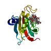
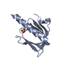
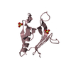
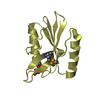
- PDB-6xcl: Crystal Structure of human telomeric DNA G-quadruplex in complex ... -
+ Open data

- Basic information
| Entry | Database: PDB / ID: 6xcl | ||||||||||||||||||||||||||||
|---|---|---|---|---|---|---|---|---|---|---|---|---|---|---|---|---|---|---|---|---|---|---|---|---|---|---|---|---|---|
| Title | Crystal Structure of human telomeric DNA G-quadruplex in complex with a novel platinum(II) complex. | ||||||||||||||||||||||||||||
 Components Components | DNA (5'-D(* Keywords KeywordsDNA / guanine quadruplex / Ligand Complex | Function / homology | : / Chem-V4A / DNA / DNA (> 10) |  Function and homology information Function and homology informationBiological species |  Homo sapiens (human) Homo sapiens (human)Method |  X-RAY DIFFRACTION / SYNCHROTRON / MOLECULAR REPLACEMENT / Resolution: 2.7 Å X-RAY DIFFRACTION / SYNCHROTRON / MOLECULAR REPLACEMENT / Resolution: 2.7 Å  Authors AuthorsMiron, C.E. / van Staalduinen, L.M. / Jia, Z. / Petitjean, A. | Funding support | |  Canada, 1items Canada, 1items
 CitationJournal: Angew.Chem.Int.Ed.Engl. / Year: 2021 CitationJournal: Angew.Chem.Int.Ed.Engl. / Year: 2021Title: Going Platinum to the Tune of a Remarkable Guanine Quadruplex Binder: Solution- and Solid-State Investigations. Authors: Miron, C.E. / van Staalduinen, L. / Rangaswamy, A.M. / Chen, M. / Liang, Y. / Jia, Z. / Mergny, J.L. / Petitjean, A. History |
|
- Structure visualization
| Structure viewer | Molecule: MolmilJmol/JSmol |
|---|
- Downloads & links
-Download
| PDBx/mmCIF format | 6xcl.cif.gz | 45.7 KB | Display | PDBx/mmCIF format |
|---|---|---|---|---|
| PDB format | pdb6xcl.ent.gz | 26.9 KB | Display | PDB format |
| PDBx/mmJSON format | 6xcl.json.gz | Tree view | PDBx/mmJSON format | |
| Others |  Other downloads Other downloads |
-Validation report
| Summary document | 6xcl_validation.pdf.gz | 1.3 MB | Display | wwPDB validaton report |
|---|---|---|---|---|
| Full document | 6xcl_full_validation.pdf.gz | 1.3 MB | Display | |
| Data in XML | 6xcl_validation.xml.gz | 4.1 KB | Display | |
| Data in CIF | 6xcl_validation.cif.gz | 5.1 KB | Display | |
| Arichive directory | https://data.pdbj.org/pub/pdb/validation_reports/xc/6xclftp://data.pdbj.org/pub/pdb/validation_reports/xc/6xcl | HTTPS FTP |
-Related structure data
| Related structure data |  3t5eS S: Starting model for refinement |
|---|---|
| Similar structure data |

-Links
 PDBj
PDBj


- Assembly
| Deposited unit | 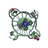
| ||||||||||||
|---|---|---|---|---|---|---|---|---|---|---|---|---|---|
| 1 |
| ||||||||||||
| Unit cell |
|
-Components
| #1: DNA chain | Mass: 6983.497 Da / Num. of mol.: 2 / Source method: obtained synthetically / Source: (synth.) Homo sapiens (human)#2: Chemical | 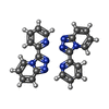  Mass: 587.494 Da / Num. of mol.: 3 / Source method: obtained synthetically / Formula: C22H16N8Pt / Feature type: SUBJECT OF INVESTIGATION Mass: 587.494 Da / Num. of mol.: 3 / Source method: obtained synthetically / Formula: C22H16N8Pt / Feature type: SUBJECT OF INVESTIGATION#3: Chemical |   Mass: 62.068 Da / Num. of mol.: 2 / Source method: obtained synthetically / Formula: C2H6O2 Mass: 62.068 Da / Num. of mol.: 2 / Source method: obtained synthetically / Formula: C2H6O2#4: Chemical | ChemComp-K /   Mass: 39.098 Da / Num. of mol.: 5 / Source method: obtained synthetically / Formula: K Mass: 39.098 Da / Num. of mol.: 5 / Source method: obtained synthetically / Formula: K#5: Water | ChemComp-HOH / |  Mass: 18.015 Da / Num. of mol.: 6 / Source method: isolated from a natural source / Formula: H2O Mass: 18.015 Da / Num. of mol.: 6 / Source method: isolated from a natural source / Formula: H2OHas ligand of interest | Y | |
|---|
-Experimental details
-Experiment
| Experiment | Method: X-RAY DIFFRACTION / Number of used crystals: 1 |
|---|
- Sample preparation
| Crystal | Density Matthews: 3.58 Å3/Da / Density % sol: 65.63 % |
|---|---|
| Crystal grow | Temperature: 285 K / Method: vapor diffusion, hanging drop / pH: 6 Details: 100 mM KCl, 10 mM MgCl2, 50 mM MES (pH 6.0), 10% PEG 400 |
-Data collection
| Diffraction | Mean temperature: 100 K / Serial crystal experiment: N |
|---|---|
| Diffraction source | Source: SYNCHROTRON / Site: CLSI / Beamline: 08B1-1 / Wavelength: 1.0332 Å |
| Detector | Type: MARMOSAIC 300 mm CCD / Detector: CCD / Date: Feb 7, 2019 |
| Radiation | Protocol: SINGLE WAVELENGTH / Monochromatic (M) / Laue (L): M / Scattering type: x-ray |
| Radiation wavelength | Wavelength: 1.0332 Å / Relative weight: 1 |
| Reflection | Resolution: 2.7→35 Å / Num. obs: 10637 / % possible obs: 99.8 % / Redundancy: 5.8 % / Biso Wilson estimate: 55.79 Å2 / Rsym value: 0.097 / Net I/σ(I): 14.28 |
| Reflection shell | Resolution: 2.7→2.9 Å / Redundancy: 5.8 % / Mean I/σ(I) obs: 2.51 / Num. unique obs: 2054 / Rsym value: 0.816 / % possible all: 99.7 |
- Processing
| Software |
| |||||||||||||||||||||||||||||||||||
|---|---|---|---|---|---|---|---|---|---|---|---|---|---|---|---|---|---|---|---|---|---|---|---|---|---|---|---|---|---|---|---|---|---|---|---|---|
| Refinement | Method to determine structure: MOLECULAR REPLACEMENT Starting model: 3t5e Resolution: 2.7→33.6 Å / SU ML: 0.3274 / Cross valid method: FREE R-VALUE / σ(F): 1.39 / Phase error: 27.956 Stereochemistry target values: GeoStd + Monomer Library + CDL v1.2
| |||||||||||||||||||||||||||||||||||
| Solvent computation | Shrinkage radii: 0.9 Å / VDW probe radii: 1.11 Å / Solvent model: FLAT BULK SOLVENT MODEL | |||||||||||||||||||||||||||||||||||
| Displacement parameters | Biso mean: 41.07 Å2 | |||||||||||||||||||||||||||||||||||
| Refinement step | Cycle: LAST / Resolution: 2.7→33.6 Å
| |||||||||||||||||||||||||||||||||||
| Refine LS restraints |
| |||||||||||||||||||||||||||||||||||
| LS refinement shell |
|
