 Movie
Movie Controller

[English] 日本語
 Yorodumi
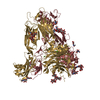
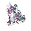
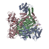
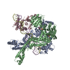
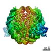
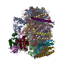
- PDB-6wxu: CryoEM structure of mouse DUOX1-DUOXA1 complex in the dimer-of-di... -
+ Open data

- Basic information
| Entry | Database: PDB / ID: 6wxu | |||||||||||||||||||||||||||
|---|---|---|---|---|---|---|---|---|---|---|---|---|---|---|---|---|---|---|---|---|---|---|---|---|---|---|---|---|
| Title | CryoEM structure of mouse DUOX1-DUOXA1 complex in the dimer-of-dimer state | |||||||||||||||||||||||||||
 Components Components | (Dual oxidase ...) x 2 | |||||||||||||||||||||||||||
 Keywords Keywords | MEMBRANE PROTEIN / protein complex / NADPH oxidase / ROS production | |||||||||||||||||||||||||||
| Function / homology |  Function and homology information Function and homology informationregulation of thyroid hormone generation / Thyroxine biosynthesis / cuticle development / NAD(P)H oxidase (H2O2-forming) / positive regulation of hydrogen peroxide biosynthetic process / hormone biosynthetic process / hydrogen peroxide metabolic process / NAD(P)H oxidase H2O2-forming activity / superoxide-generating NAD(P)H oxidase activity / NADPH oxidase complex ...regulation of thyroid hormone generation / Thyroxine biosynthesis / cuticle development / NAD(P)H oxidase (H2O2-forming) / positive regulation of hydrogen peroxide biosynthetic process / hormone biosynthetic process / hydrogen peroxide metabolic process / NAD(P)H oxidase H2O2-forming activity / superoxide-generating NAD(P)H oxidase activity / NADPH oxidase complex / thyroid hormone generation / Oxidoreductases / hydrogen peroxide biosynthetic process / superoxide anion generation / positive regulation of cell motility / positive regulation of wound healing / cell leading edge / Oxidoreductases; Acting on a peroxide as acceptor; Peroxidases / response to cAMP / NADPH binding / FAD binding / positive regulation of neuron differentiation / reactive oxygen species metabolic process / hydrogen peroxide catabolic process / peroxidase activity / defense response / positive regulation of reactive oxygen species metabolic process / cytokine-mediated signaling pathway / intracellular protein localization / protein transport / regulation of inflammatory response / response to oxidative stress / apical plasma membrane / protein heterodimerization activity / heme binding / calcium ion binding / endoplasmic reticulum membrane / enzyme binding / cell surface / endoplasmic reticulum / membrane / plasma membrane Similarity search - Function | |||||||||||||||||||||||||||
| Biological species |  | |||||||||||||||||||||||||||
| Method | ELECTRON MICROSCOPY / single particle reconstruction / cryo EM / Resolution: 2.7 Å | |||||||||||||||||||||||||||
 Authors Authors | Sun, J. | |||||||||||||||||||||||||||
| Funding support |  United States, 1items United States, 1items
| |||||||||||||||||||||||||||
 Citation Citation | Journal: Nat Struct Mol Biol / Year: 2020 Title: Structures of mouse DUOX1-DUOXA1 provide mechanistic insights into enzyme activation and regulation. Authors: Ji Sun / Abstract: DUOX1, an NADPH oxidase family member, catalyzes the production of hydrogen peroxide. DUOX1 is expressed in various tissues, including the thyroid and respiratory tract, and plays a crucial role in ...DUOX1, an NADPH oxidase family member, catalyzes the production of hydrogen peroxide. DUOX1 is expressed in various tissues, including the thyroid and respiratory tract, and plays a crucial role in processes such as thyroid hormone biosynthesis and innate host defense. DUOX1 co-assembles with its maturation factor DUOXA1 to form an active enzyme complex. However, the molecular mechanisms for activation and regulation of DUOX1 remain mostly unclear. Here, I present cryo-EM structures of the mammalian DUOX1-DUOXA1 complex, in the absence and presence of substrate NADPH, as well as DUOX1-DUOXA1 in an unexpected dimer-of-dimers configuration. These structures reveal atomic details of the DUOX1-DUOXA1 interaction, a lipid-mediated NADPH-binding pocket and the electron transfer path. Furthermore, biochemical and structural analyses indicate that the dimer-of-dimers configuration represents an inactive state of DUOX1-DUOXA1, suggesting an oligomerization-dependent regulatory mechanism. Together, my work provides structural bases for DUOX1-DUOXA1 activation and regulation. | |||||||||||||||||||||||||||
| History |
|
- Structure visualization
| Movie |
Movie viewer |
|---|---|
| Structure viewer | Molecule: MolmilJmol/JSmol |
- Downloads & links
-Download
| PDBx/mmCIF format | 6wxu.cif.gz | 444.6 KB | Display | PDBx/mmCIF format |
|---|---|---|---|---|
| PDB format | pdb6wxu.ent.gz | 337.6 KB | Display | PDB format |
| PDBx/mmJSON format | 6wxu.json.gz | Tree view | PDBx/mmJSON format | |
| Others |  Other downloads Other downloads |
-Validation report
| Arichive directory | https://data.pdbj.org/pub/pdb/validation_reports/wx/6wxuftp://data.pdbj.org/pub/pdb/validation_reports/wx/6wxu | HTTPS FTP |
|---|
-Related structure data
| Related structure data |  21963MC  6wxrC  6wxvC M: map data used to model this data C: citing same article ( |
|---|---|
| Similar structure data |


-Links
 PDBj
PDBj


- Assembly
| Deposited unit | 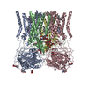
|
|---|---|
| 1 |
|
-Components
-Dual oxidase ... , 2 types, 4 molecules ACBD
| #1: Protein | Mass: 175739.812 Da / Num. of mol.: 2 Source method: isolated from a genetically manipulated source Source: (gene. exp.)  Homo sapiens (human) / References: UniProt: A2AQ92 Homo sapiens (human) / References: UniProt: A2AQ92#2: Protein | Mass: 37619.738 Da / Num. of mol.: 2 Source method: isolated from a genetically manipulated source Source: (gene. exp.) Homo sapiens (human) / References: UniProt: Q8VE49 |
|---|
-Sugars , 2 types, 12 molecules 
| #3: Polysaccharide | Source method: isolated from a genetically manipulated source #4: Sugar | ChemComp-NAG /  Type: D-saccharide, beta linking / Mass: 221.208 Da / Num. of mol.: 10 / Source method: obtained synthetically / Formula: C8H15NO6 Type: D-saccharide, beta linking / Mass: 221.208 Da / Num. of mol.: 10 / Source method: obtained synthetically / Formula: C8H15NO6 |
|---|
-Non-polymers , 3 types, 8 molecules 


| #5: Chemical | ChemComp-HEC /  Mass: 618.503 Da / Num. of mol.: 4 / Source method: obtained synthetically / Formula: C34H34FeN4O4 / Feature type: SUBJECT OF INVESTIGATION Mass: 618.503 Da / Num. of mol.: 4 / Source method: obtained synthetically / Formula: C34H34FeN4O4 / Feature type: SUBJECT OF INVESTIGATION#6: Chemical |  Mass: 535.671 Da / Num. of mol.: 2 Mass: 535.671 Da / Num. of mol.: 2Source method: isolated from a genetically manipulated source Formula: C27H52O8P #7: Chemical |  Mass: 622.834 Da / Num. of mol.: 2 Mass: 622.834 Da / Num. of mol.: 2Source method: isolated from a genetically manipulated source Formula: C32H65NO8P / Comment: phospholipid*YM |
|---|
-Details
| Has ligand of interest | Y |
|---|---|
| Has protein modification | Y |
-Experimental details
-Experiment
| Experiment | Method: ELECTRON MICROSCOPY |
|---|---|
| EM experiment | Aggregation state: PARTICLE / 3D reconstruction method: single particle reconstruction |
- Sample preparation
| Component | Name: mouse DUOX1-DUOXA1 complex / Type: COMPLEX / Entity ID: #1-#2 / Source: RECOMBINANT |
|---|---|
| Molecular weight | Value: 0.22 MDa / Experimental value: NO |
| Source (natural) | Organism: |
| Source (recombinant) | Organism: Homo sapiens (human) |
| Buffer solution | pH: 8 |
| Specimen | Embedding applied: NO / Shadowing applied: NO / Staining applied: NO / Vitrification applied: YES |
| Vitrification | Cryogen name: ETHANE / Humidity: 100 % |
- Electron microscopy imaging
| Experimental equipment |  Model: Titan Krios / Image courtesy: FEI Company |
|---|---|
| Microscopy | Model: FEI TITAN KRIOS |
| Electron gun | Electron source:  FIELD EMISSION GUN / Accelerating voltage: 300 kV / Illumination mode: FLOOD BEAM FIELD EMISSION GUN / Accelerating voltage: 300 kV / Illumination mode: FLOOD BEAM |
| Electron lens | Mode: BRIGHT FIELD |
| Image recording | Electron dose: 65.4 e/Å2 / Film or detector model: GATAN K3 (6k x 4k) |
- Processing
| Software | Name: PHENIX / Version: 1.17.1_3660: / Classification: refinement | ||||||||||||||||||||||||
|---|---|---|---|---|---|---|---|---|---|---|---|---|---|---|---|---|---|---|---|---|---|---|---|---|---|
| EM software |
| ||||||||||||||||||||||||
| CTF correction | Type: NONE | ||||||||||||||||||||||||
| Symmetry | Point symmetry: C2 (2 fold cyclic) | ||||||||||||||||||||||||
| 3D reconstruction | Resolution: 2.7 Å / Resolution method: FSC 0.143 CUT-OFF / Num. of particles: 302097 / Symmetry type: POINT | ||||||||||||||||||||||||
| Atomic model building | Protocol: AB INITIO MODEL | ||||||||||||||||||||||||
| Refine LS restraints |
|
