 Movie
Movie Controller

[English] 日本語
 Yorodumi
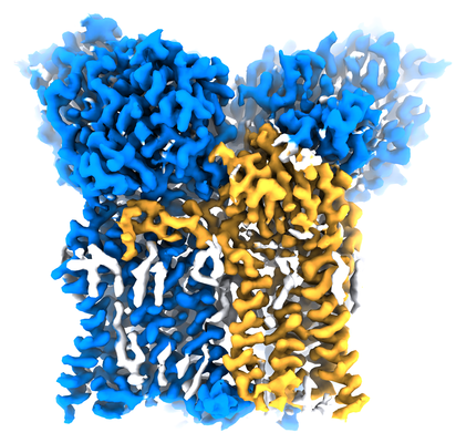
- EMDB-21963: CryoEM structure of mouse DUOX1-DUOXA1 complex in the dimer-of-di... -
+ Open data

- Basic information
| Entry | Database: EMDB / ID: EMD-21963 | |||||||||
|---|---|---|---|---|---|---|---|---|---|---|
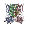
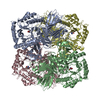
| Title | CryoEM structure of mouse DUOX1-DUOXA1 complex in the dimer-of-dimer state | |||||||||
 Map data Map data | cryoEM structure of the mouse DUOX1-DUOXA1 complex in the dimer-of-dimer state | |||||||||
 Sample Sample |
| |||||||||
 Keywords Keywords | membrane protein / protein complex / NADPH oxidase / ROS production | |||||||||
| Function / homology |  Function and homology information Function and homology informationregulation of thyroid hormone generation / Thyroxine biosynthesis / cuticle development / NAD(P)H oxidase (H2O2-forming) / positive regulation of hydrogen peroxide biosynthetic process / hormone biosynthetic process / hydrogen peroxide metabolic process / NAD(P)H oxidase H2O2-forming activity / superoxide-generating NAD(P)H oxidase activity / NADPH oxidase complex ...regulation of thyroid hormone generation / Thyroxine biosynthesis / cuticle development / NAD(P)H oxidase (H2O2-forming) / positive regulation of hydrogen peroxide biosynthetic process / hormone biosynthetic process / hydrogen peroxide metabolic process / NAD(P)H oxidase H2O2-forming activity / superoxide-generating NAD(P)H oxidase activity / NADPH oxidase complex / thyroid hormone generation / Oxidoreductases / hydrogen peroxide biosynthetic process / superoxide anion generation / positive regulation of cell motility / positive regulation of wound healing / cell leading edge / Oxidoreductases; Acting on a peroxide as acceptor; Peroxidases / response to cAMP / NADPH binding / FAD binding / positive regulation of neuron differentiation / reactive oxygen species metabolic process / hydrogen peroxide catabolic process / peroxidase activity / defense response / positive regulation of reactive oxygen species metabolic process / cytokine-mediated signaling pathway / intracellular protein localization / protein transport / regulation of inflammatory response / response to oxidative stress / apical plasma membrane / protein heterodimerization activity / heme binding / calcium ion binding / endoplasmic reticulum membrane / enzyme binding / cell surface / endoplasmic reticulum / membrane / plasma membrane Similarity search - Function | |||||||||
| Biological species |  | |||||||||
| Method | single particle reconstruction / cryo EM / Resolution: 2.7 Å | |||||||||
 Authors Authors | Sun J | |||||||||
| Funding support |  United States, 1 items United States, 1 items
| |||||||||
 Citation Citation | Journal: Nat Struct Mol Biol / Year: 2020 Title: Structures of mouse DUOX1-DUOXA1 provide mechanistic insights into enzyme activation and regulation. Authors: Ji Sun / Abstract: DUOX1, an NADPH oxidase family member, catalyzes the production of hydrogen peroxide. DUOX1 is expressed in various tissues, including the thyroid and respiratory tract, and plays a crucial role in ...DUOX1, an NADPH oxidase family member, catalyzes the production of hydrogen peroxide. DUOX1 is expressed in various tissues, including the thyroid and respiratory tract, and plays a crucial role in processes such as thyroid hormone biosynthesis and innate host defense. DUOX1 co-assembles with its maturation factor DUOXA1 to form an active enzyme complex. However, the molecular mechanisms for activation and regulation of DUOX1 remain mostly unclear. Here, I present cryo-EM structures of the mammalian DUOX1-DUOXA1 complex, in the absence and presence of substrate NADPH, as well as DUOX1-DUOXA1 in an unexpected dimer-of-dimers configuration. These structures reveal atomic details of the DUOX1-DUOXA1 interaction, a lipid-mediated NADPH-binding pocket and the electron transfer path. Furthermore, biochemical and structural analyses indicate that the dimer-of-dimers configuration represents an inactive state of DUOX1-DUOXA1, suggesting an oligomerization-dependent regulatory mechanism. Together, my work provides structural bases for DUOX1-DUOXA1 activation and regulation. | |||||||||
| History |
|
- Structure visualization
| Movie |
Movie viewer |
|---|---|
| Structure viewer | EM map: SurfViewMolmilJmol/JSmol |
| Supplemental images |
- Downloads & links
-EMDB archive
| Map data | emd_21963.map.gz | 43.7 MB | EMDB map data format | |
|---|---|---|---|---|
| Header (meta data) | emd-21963-v30.xmlemd-21963.xml | 16.6 KB 16.6 KB | Display Display | EMDB header |
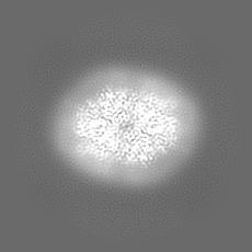
| Images |  emd_21963.png emd_21963.png | 210.7 KB | ||
| Filedesc metadata | emd-21963.cif.gz | 7.3 KB | ||
| Archive directory |  http://ftp.pdbj.org/pub/emdb/structures/EMD-21963ftp://ftp.pdbj.org/pub/emdb/structures/EMD-21963 http://ftp.pdbj.org/pub/emdb/structures/EMD-21963ftp://ftp.pdbj.org/pub/emdb/structures/EMD-21963 | HTTPS FTP |
-Related structure data
| Related structure data |  6wxuMC  6wxrC  6wxvC M: atomic model generated by this map C: citing same article ( |
|---|---|
| Similar structure data |


-Links
| EMDB pages | EMDB (EBI/PDBe) / EMDataResource |
|---|---|
| Related items in Molecule of the Month |

-Map
| File | Download / File: emd_21963.map.gz / Format: CCP4 / Size: 46.4 MB / Type: IMAGE STORED AS FLOATING POINT NUMBER (4 BYTES) | ||||||||||||||||||||||||||||||||||||||||||||||||||||||||||||||||||||
|---|---|---|---|---|---|---|---|---|---|---|---|---|---|---|---|---|---|---|---|---|---|---|---|---|---|---|---|---|---|---|---|---|---|---|---|---|---|---|---|---|---|---|---|---|---|---|---|---|---|---|---|---|---|---|---|---|---|---|---|---|---|---|---|---|---|---|---|---|---|
| Annotation | cryoEM structure of the mouse DUOX1-DUOXA1 complex in the dimer-of-dimer state | ||||||||||||||||||||||||||||||||||||||||||||||||||||||||||||||||||||
| Projections & slices | Image control
Images are generated by Spider. | ||||||||||||||||||||||||||||||||||||||||||||||||||||||||||||||||||||
| Voxel size | X=Y=Z: 1.08 Å | ||||||||||||||||||||||||||||||||||||||||||||||||||||||||||||||||||||
| Density |
| ||||||||||||||||||||||||||||||||||||||||||||||||||||||||||||||||||||
| Symmetry | Space group: 1 | ||||||||||||||||||||||||||||||||||||||||||||||||||||||||||||||||||||
| Details | EMDB XML:
CCP4 map header:
| ||||||||||||||||||||||||||||||||||||||||||||||||||||||||||||||||||||
 Z (Sec.)
 Y (Row.)
 X (Col.)


-Supplemental data
- Sample components
-Entire : mouse DUOX1-DUOXA1 complex
| Entire | Name: mouse DUOX1-DUOXA1 complex |
|---|---|
| Components |
|
-Supramolecule #1: mouse DUOX1-DUOXA1 complex
| Supramolecule | Name: mouse DUOX1-DUOXA1 complex / type: complex / ID: 1 / Parent: 0 / Macromolecule list: #1-#2 |
|---|---|
| Source (natural) | Organism: |
| Molecular weight | Theoretical: 220 KDa |
-Macromolecule #1: Dual oxidase 1
| Macromolecule | Name: Dual oxidase 1 / type: protein_or_peptide / ID: 1 / Number of copies: 2 / Enantiomer: LEVO |
|---|---|
| Source (natural) | Organism: |
| Molecular weight | Theoretical: 175.739812 KDa |
| Recombinant expression | Organism:  Homo sapiens (human) Homo sapiens (human) |
| Sequence | String: GPSRGAQNSI SWEVQRFDGW YNNLMEHRWG SKGSRLQRLV PASYADGVYQ PLKEPYLPNP RHLSNRVMRG SAGQPSLRNR TVLGVFFGY HVLSDLVSVE TPGCPAEFLN IYIPHGDPVF DPDKRGNVVL PFQRSRWDRN TGQSPSNPRD QSNQVTGWLD G SAIYGSSH ...String: GPSRGAQNSI SWEVQRFDGW YNNLMEHRWG SKGSRLQRLV PASYADGVYQ PLKEPYLPNP RHLSNRVMRG SAGQPSLRNR TVLGVFFGY HVLSDLVSVE TPGCPAEFLN IYIPHGDPVF DPDKRGNVVL PFQRSRWDRN TGQSPSNPRD QSNQVTGWLD G SAIYGSSH SWSDTLRSFS GGQLASGPDP AFPSDSQSSL LMWMAPDPST GQGGPRGVYA FGAQRGNREP FLQALGLLWF RY HNLCARK LAQEHPHWGD EELFQHARKR VIATYQNIAM YEWLPSFLKQ TPPEYPGYRP FLDPSISPEF VVASEQFLST MVP SGVYMR NASCHFQGIP SHNSSVSGAL RVCNSYWSRE HPKLQRAEDV DALLLGMASQ IAEREDHVVV EDMQDFWPGP LKFS RTDYL ASCLQRGRDL GLPSYTKARE ALGLSPISHW QDINPALSRS NGTVLEATAA LYNQDLSRLE LLPGGLLESH GDPGP LFST IVLDQFVRLR DGDRYWFENT RNGLFSKEEI AEIRNTSLRD ILVAVTNVDP SALQPNVFFW LAGDPCPQPS QLSAKG LPA CAPLFIRDYF EGSGFGFGLT IGTLCCFPLV SLLSAWIVAR LRKRNFKRLQ RQDRQSIMSE KLVGGVEALE WQGRNEP CR PVLVHLQPGQ IRVVDGRLTV LRTIQLRPPQ QVNLILSSNR GRRTLLLKIP KEYDLVLLFN MEEERQALVE NVRGALKE N GLSFQEWELR EQELMRAAVT RQQRGHLLET FFRHLFSQVL DINQADAGTL PLDSSTKVRE ALTCELSRAE FADSLGLKP QDMFVESMFS LADKDGNGYL SFREFLDILV VFMKGSPEEK SRLMFRMYDF DGNGLISKDE FIRMLRSFIE ISNNCLSKAQ LAEVVESMF RESGFQDKEE LTWEDFHFML RDHDSDLRFT QLCVKGVEVP EVIKNLCRRA SYISQEKICP SPRMSAHCAR N NMKTASSP QRLQCPMDTD PPQEIRRRFG KKVTSFQPLL FTEAHREKFQ RSRRHQTVQQ FKRFIENYRR HIGCVAVFYT IT GALFLER AYYYAFAAHH SGITDTTRVG IILSRGTAAS ISFMFSYILL TMCRNLITFL RETFLNRYIP FDAAVDFHRL IAS TAIILT VLHSAGHVVN VYLFSISPLS VLSCLFPGLF HDDGSEFPQK YYWWFFQTVP GLTGVLLLLA LAIMYVFASH HFRR RSFRG FWLTHHLYIF LYILLIIHGS FALIQMPRFH IFFLVPAIIY VGDKLVSLSR KKVEISVVKA ELLPSGVTHL RFQRP QGFE YKSGQWVRIA CLALGTTEYH PFTLTSAPHE DTLSLHIRAA GPWTTRLREI YSPPTGDTCA RYPKLYLDGP FGEGHQ EWH KFEVSVLVGG GIGVTPFASI LKDLVFKSSV SCQVFCKKIY FIWVTRTQRQ FEWLADIIRE VEENDRQDLV SVHIYIT QL AEKFDLRTTM LYICERHFQK VLNRSLFTGL RSITHFGRPP FEPFFNSLQE VHPQVRKIGV FSCGPPGMTK NVEKACQL I NRQDRTHFSH HYENF UniProtKB: Dual oxidase 1 |
-Macromolecule #2: Dual oxidase maturation factor 1
| Macromolecule | Name: Dual oxidase maturation factor 1 / type: protein_or_peptide / ID: 2 / Number of copies: 2 / Enantiomer: LEVO |
|---|---|
| Source (natural) | Organism: |
| Molecular weight | Theoretical: 37.619738 KDa |
| Recombinant expression | Organism: Homo sapiens (human) |
| Sequence | String: MAALGHTLPF YTGTKPTFPM DTTLAVIITI FLTALVTFII ILPGIRGKTR LFWLLRVVTS LFIGAVILAV NFSSEWSVGH VNANTTYKA FSPKWVSVDV GLQIGLGGVN ITLTGTPVQQ LNETINYNEA FAWRLGRSYA EEYAKALEKG LPDPVLYLAE K FTPRSPCG ...String: MAALGHTLPF YTGTKPTFPM DTTLAVIITI FLTALVTFII ILPGIRGKTR LFWLLRVVTS LFIGAVILAV NFSSEWSVGH VNANTTYKA FSPKWVSVDV GLQIGLGGVN ITLTGTPVQQ LNETINYNEA FAWRLGRSYA EEYAKALEKG LPDPVLYLAE K FTPRSPCG LYNQYRLAGH YASAMLWVAF LCWLLANVML SMPVLVYGGH MLLATGLFQL LALFFFSMTT SLISPCPLRL GT AVLHTHH GPAFWITLAT GLLCILLGLV MAVAHRMQPH RLKAFFNQSS EDPVLEWGSE EGGLLSPHYR SIAESPETQD IPM SVASSE TCFKEEHPKE SDCSL UniProtKB: Dual oxidase maturation factor 1 |
-Macromolecule #4: 2-acetamido-2-deoxy-beta-D-glucopyranose
| Macromolecule | Name: 2-acetamido-2-deoxy-beta-D-glucopyranose / type: ligand / ID: 4 / Number of copies: 10 / Formula: NAG |
|---|---|
| Molecular weight | Theoretical: 221.208 Da |
| Chemical component information |  ChemComp-NAG: |
-Macromolecule #5: HEME C
| Macromolecule | Name: HEME C / type: ligand / ID: 5 / Number of copies: 4 / Formula: HEC |
|---|---|
| Molecular weight | Theoretical: 618.503 Da |
| Chemical component information |  ChemComp-HEC: |
-Macromolecule #6: 1,2-DILAUROYL-SN-GLYCERO-3-PHOSPHATE
| Macromolecule | Name: 1,2-DILAUROYL-SN-GLYCERO-3-PHOSPHATE / type: ligand / ID: 6 / Number of copies: 2 / Formula: PX2 |
|---|---|
| Molecular weight | Theoretical: 535.671 Da |
| Chemical component information |  ChemComp-PX2: |
-Macromolecule #7: DIUNDECYL PHOSPHATIDYL CHOLINE
| Macromolecule | Name: DIUNDECYL PHOSPHATIDYL CHOLINE / type: ligand / ID: 7 / Number of copies: 2 / Formula: PLC |
|---|---|
| Molecular weight | Theoretical: 622.834 Da |
| Chemical component information |  ChemComp-PLC: |
-Experimental details
-Structure determination
| Method | cryo EM |
|---|---|
 Processing Processing | single particle reconstruction |
| Aggregation state | particle |
-Sample preparation
| Buffer | pH: 8 |
|---|---|
| Vitrification | Cryogen name: ETHANE / Chamber humidity: 100 % |
- Electron microscopy
| Microscope | FEI TITAN KRIOS |
|---|---|
| Image recording | Film or detector model: GATAN K3 (6k x 4k) / Average electron dose: 65.4 e/Å2 |
| Electron beam | Acceleration voltage: 300 kV / Electron source:  FIELD EMISSION GUN FIELD EMISSION GUN |
| Electron optics | Illumination mode: FLOOD BEAM / Imaging mode: BRIGHT FIELD |
| Experimental equipment |  Model: Titan Krios / Image courtesy: FEI Company |
+Image processing
-Atomic model buiding 1
| Refinement | Protocol: AB INITIO MODEL |
|---|---|
| Output model | PDB-6wxu: |
