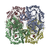
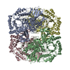
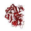
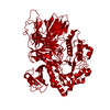
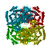
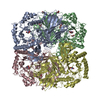
Crystal structure of GH36 alpha-galactosidase AgaA A355E D478A from Geobacillus stearothermophilus in complex with stachyose
Components
Alpha-galactosidase AgaA
Keywords
HYDROLASE / Glycoside Hydrolase / carbohydrate
Function / homology
Function and homology information
stachyose metabolic process / raffinose catabolic process / alpha-galactosidase / alpha-galactosidase activity / protein homotetramerization Similarity search - Function
alpha-galactosidase from lactobacil brevis / Glycosyl hydrolase family 36, N-terminal / Glycosyl hydrolase family 36, C-terminal / Alpha-galactosidase, N-terminal domain superfamily / Glycosyl hydrolase family 36 C-terminal domain / Glycosyl hydrolase family 36 N-terminal domain / Glycoside hydrolase family 36 / Melibiase / : / Glycoside hydrolase family 27/36, conserved site ...alpha-galactosidase from lactobacil brevis / Glycosyl hydrolase family 36, N-terminal / Glycosyl hydrolase family 36, C-terminal / Alpha-galactosidase, N-terminal domain superfamily / Glycosyl hydrolase family 36 C-terminal domain / Glycosyl hydrolase family 36 N-terminal domain / Glycoside hydrolase family 36 / Melibiase / : / Glycoside hydrolase family 27/36, conserved site / Alpha-galactosidase signature. / Beta-galactosidase; Chain A, domain 5 / Golgi alpha-mannosidase II / Glycosyl hydrolase, all-beta / Distorted Sandwich / Aldolase class I / Aldolase-type TIM barrel / Glycoside hydrolase superfamily / TIM Barrel / Alpha-Beta Barrel / Immunoglobulin-like / Sandwich / Mainly Beta / Alpha Beta Similarity search - Domain/homology
In the structure databanks used in Yorodumi, some data are registered as the other names, "COVID-19 virus" and "2019-nCoV". Here are the details of the virus and the list of structure data.
Jan 31, 2019. EMDB accession codes are about to change! (news from PDBe EMDB page)
EMDB accession codes are about to change! (news from PDBe EMDB page)
The allocation of 4 digits for EMDB accession codes will soon come to an end. Whilst these codes will remain in use, new EMDB accession codes will include an additional digit and will expand incrementally as the available range of codes is exhausted. The current 4-digit format prefixed with “EMD-” (i.e. EMD-XXXX) will advance to a 5-digit format (i.e. EMD-XXXXX), and so on. It is currently estimated that the 4-digit codes will be depleted around Spring 2019, at which point the 5-digit format will come into force.
The EM Navigator/Yorodumi systems omit the EMD- prefix.
Related info.:Q: What is EMD? / ID/Accession-code notation in Yorodumi/EM Navigator
Yorodumi is a browser for structure data from EMDB, PDB, SASBDB, etc.
This page is also the successor to EM Navigator detail page, and also detail information page/front-end page for Omokage search.
The word "yorodu" (or yorozu) is an old Japanese word meaning "ten thousand". "mi" (miru) is to see.
Related info.:EMDB / PDB / SASBDB / Comparison of 3 databanks / Yorodumi Search / Aug 31, 2016. New EM Navigator & Yorodumi / Yorodumi Papers / Jmol/JSmol / Function and homology information / Changes in new EM Navigator and Yorodumi
 Movie
Movie Controller

 Yorodumi
 Open data
 Basic information
 Components
 Keywords
 Function and homology information
 Geobacillus stearothermophilus (bacteria)
 X-RAY DIFFRACTION /
  Authors
 Citation
 Structure visualization
 Downloads & links
 Other downloads
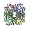


 PDBj
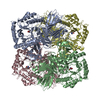
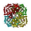
PDBj Assembly
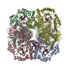
 Sample preparation
 / Beamline: X06DA / Wavelength: 1
  Processing