 Movie
Movie Controller

[English] 日本語
 Yorodumi
- PDB-2xn2: Structure of alpha-galactosidase from Lactobacillus acidophilus N... -
+ Open data

- Basic information
| Entry | Database: PDB / ID: 2xn2 | ||||||
|---|---|---|---|---|---|---|---|
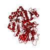
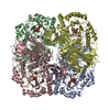
| Title | Structure of alpha-galactosidase from Lactobacillus acidophilus NCFM with galactose | ||||||
 Components Components | ALPHA-GALACTOSIDASE | ||||||
 Keywords Keywords | HYDROLASE / GLYCOSIDASE | ||||||
| Function / homology |  Function and homology information Function and homology informationresponse to raffinose / alpha-galactosidase / alpha-galactosidase activity / carbohydrate catabolic process / protein homotetramerization Similarity search - Function | ||||||
| Biological species |  LACTOBACILLUS ACIDOPHILUS NCFM (bacteria) LACTOBACILLUS ACIDOPHILUS NCFM (bacteria) | ||||||
| Method |  X-RAY DIFFRACTION / SYNCHROTRON / MOLECULAR REPLACEMENT / Resolution: 1.58 Å X-RAY DIFFRACTION / SYNCHROTRON / MOLECULAR REPLACEMENT / Resolution: 1.58 Å | ||||||
 Authors Authors | Fredslund, F. / Abou Hachem, M. / Larsen, R.J. / Sorensen, P.G. / Lo Leggio, L. / Svensson, B. | ||||||
 Citation Citation | Journal: J.Mol.Biol. / Year: 2011 Title: Crystal Structure of Alpha-Galactosidase from Lactobacillus Acidophilus Ncfm: Insight Into Tetramer Formation and Substrate Binding. Authors: Fredslund, F. / Abou Hachem, M. / Jonsgaard Larsen, R. / Gerd Sorensen, P. / Coutinho, P.M. / Lo Leggio, L. / Svensson, B. | ||||||
| History |
| ||||||
| Remark 700 | SHEET DETERMINATION METHOD: DSSP THE SHEETS PRESENTED AS "AI" IN EACH CHAIN ON SHEET RECORDS BELOW ... SHEET DETERMINATION METHOD: DSSP THE SHEETS PRESENTED AS "AI" IN EACH CHAIN ON SHEET RECORDS BELOW IS ACTUALLY AN 8-STRANDED BARREL THIS IS REPRESENTED BY A 9-STRANDED SHEET IN WHICH THE FIRST AND LAST STRANDS ARE IDENTICAL. |
- Structure visualization
| Structure viewer | Molecule: MolmilJmol/JSmol |
|---|
- Downloads & links
-Download
| PDBx/mmCIF format | 2xn2.cif.gz | 435.5 KB | Display | PDBx/mmCIF format |
|---|---|---|---|---|
| PDB format | pdb2xn2.ent.gz | 363.5 KB | Display | PDB format |
| PDBx/mmJSON format | 2xn2.json.gz | Tree view | PDBx/mmJSON format | |
| Others |  Other downloads Other downloads |
-Validation report
| Arichive directory | https://data.pdbj.org/pub/pdb/validation_reports/xn/2xn2ftp://data.pdbj.org/pub/pdb/validation_reports/xn/2xn2 | HTTPS FTP |
|---|
-Related structure data
| Related structure data |  2xn0SC  2xn1C S: Starting model for refinement C: citing same article ( |
|---|---|
| Similar structure data |


-Links
 PDBj
PDBj


- Assembly
| Deposited unit | 
| ||||||||||||
|---|---|---|---|---|---|---|---|---|---|---|---|---|---|
| 1 | 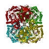
| ||||||||||||
| Unit cell |
| ||||||||||||
| Components on special symmetry positions |
|
-Components
| #1: Protein | Mass: 84619.977 Da / Num. of mol.: 1 Source method: isolated from a genetically manipulated source Source: (gene. exp.) LACTOBACILLUS ACIDOPHILUS NCFM (bacteria)Production host: References: UniProt: Q7WWP9, UniProt: G1UB44*PLUS, alpha-galactosidase | ||||
|---|---|---|---|---|---|
| #2: Sugar | ChemComp-GLA /   Type: D-saccharide, alpha linking / Mass: 180.156 Da / Num. of mol.: 1 Type: D-saccharide, alpha linking / Mass: 180.156 Da / Num. of mol.: 1Source method: isolated from a genetically manipulated source Formula: C6H12O6 | ||||
| #3: Chemical | ChemComp-IMD /   Mass: 69.085 Da / Num. of mol.: 1 / Source method: obtained synthetically / Formula: C3H5N2 Mass: 69.085 Da / Num. of mol.: 1 / Source method: obtained synthetically / Formula: C3H5N2 | ||||
| #4: Chemical | ChemComp-GOL /   Mass: 92.094 Da / Num. of mol.: 25 / Source method: obtained synthetically / Formula: C3H8O3 Mass: 92.094 Da / Num. of mol.: 25 / Source method: obtained synthetically / Formula: C3H8O3#5: Water | ChemComp-HOH / |  Mass: 18.015 Da / Num. of mol.: 624 / Source method: isolated from a natural source / Formula: H2O Mass: 18.015 Da / Num. of mol.: 624 / Source method: isolated from a natural source / Formula: H2ONonpolymer details | ALPHA D-GALACTOSE (GLA): PRESENT IN THE ACTIVE SITE IMIDAZOLE (IMD): BOUND NEAR THE ACTIVE SITE | |
-Experimental details
-Experiment
| Experiment | Method: X-RAY DIFFRACTION / Number of used crystals: 1 |
|---|
- Sample preparation
| Crystal | Density Matthews: 2.53 Å3/Da / Density % sol: 51.4 % / Description: NONE |
|---|---|
| Crystal grow | pH: 7.5 Details: 8-12% PEG8000, 0.1M IMIDAZOLE, 10% GLYCEROL, pH 7.5 |
-Data collection
| Diffraction | Mean temperature: 100 K |
|---|---|
| Diffraction source | Source: SYNCHROTRON / Site: MAX II  / Beamline: I911-2 / Wavelength: 1.03908 / Beamline: I911-2 / Wavelength: 1.03908 |
| Detector | Type: MARRESEARCH / Detector: CCD / Date: Jun 4, 2009 / Details: MIRRORS |
| Radiation | Monochromator: BENT SI (111) CRYSTAL / Protocol: SINGLE WAVELENGTH / Monochromatic (M) / Laue (L): M / Scattering type: x-ray |
| Radiation wavelength | Wavelength: 1.03908 Å / Relative weight: 1 |
| Reflection | Resolution: 1.58→19.63 Å / Num. obs: 117007 / % possible obs: 92 % / Observed criterion σ(I): 2 / Redundancy: 4.1 % / Biso Wilson estimate: 11.62 Å2 / Rmerge(I) obs: 0.09 / Net I/σ(I): 13.7 |
| Reflection shell | Resolution: 1.58→1.62 Å / Redundancy: 4 % / Rmerge(I) obs: 0.67 / Mean I/σ(I) obs: 2.53 / % possible all: 80.6 |
- Processing
| Software |
| ||||||||||||||||||||||||||||||||||||||||||||||||||||||||||||||||||||||||||||||||||||||||||||||||||||
|---|---|---|---|---|---|---|---|---|---|---|---|---|---|---|---|---|---|---|---|---|---|---|---|---|---|---|---|---|---|---|---|---|---|---|---|---|---|---|---|---|---|---|---|---|---|---|---|---|---|---|---|---|---|---|---|---|---|---|---|---|---|---|---|---|---|---|---|---|---|---|---|---|---|---|---|---|---|---|---|---|---|---|---|---|---|---|---|---|---|---|---|---|---|---|---|---|---|---|---|---|---|
| Refinement | Method to determine structure: MOLECULAR REPLACEMENT Starting model: PDB ENTRY 2XN0 Resolution: 1.58→19.632 Å / SU ML: 0.15 / σ(F): 1.99 / Phase error: 13.48 / Stereochemistry target values: ML / Details: RESIDUES 1-3 ARE DISORDERED.
| ||||||||||||||||||||||||||||||||||||||||||||||||||||||||||||||||||||||||||||||||||||||||||||||||||||
| Solvent computation | Shrinkage radii: 0.9 Å / VDW probe radii: 1.11 Å / Solvent model: FLAT BULK SOLVENT MODEL / Bsol: 55.183 Å2 / ksol: 0.473 e/Å3 | ||||||||||||||||||||||||||||||||||||||||||||||||||||||||||||||||||||||||||||||||||||||||||||||||||||
| Displacement parameters |
| ||||||||||||||||||||||||||||||||||||||||||||||||||||||||||||||||||||||||||||||||||||||||||||||||||||
| Refinement step | Cycle: LAST / Resolution: 1.58→19.632 Å
| ||||||||||||||||||||||||||||||||||||||||||||||||||||||||||||||||||||||||||||||||||||||||||||||||||||
| Refine LS restraints |
| ||||||||||||||||||||||||||||||||||||||||||||||||||||||||||||||||||||||||||||||||||||||||||||||||||||
| LS refinement shell |
| ||||||||||||||||||||||||||||||||||||||||||||||||||||||||||||||||||||||||||||||||||||||||||||||||||||
| Refinement TLS params. | Method: refined / Refine-ID: X-RAY DIFFRACTION
| ||||||||||||||||||||||||||||||||||||||||||||||||||||||||||||||||||||||||||||||||||||||||||||||||||||
| Refinement TLS group |
|
