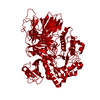
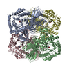
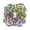
Entry Database : PDB / ID : 2yfnTitle galactosidase domain of alpha-galactosidase-sucrose kinase, AgaSK ALPHA-GALACTOSIDASE-SUCROSE KINASE AGASK Keywords Function / homology Function Domain/homology Component
/ / / / / / / / / / / / / / / / / / / / / / / / / / / / / / / / / / / / / / / / / / / / / / / / / / / / / / / Biological species RUMINOCOCCUS GNAVUS E1 (bacteria)Method / / / Resolution : 1.45 Å Authors Sulzenbacher, G. / Bruel, L. / Tison-Cervera, M. / Pujol, A. / Nicoletti, C. / Perrier, J. / Galinier, A. / Ropartz, D. / Fons, M. / Pompeo, F. / Giardina, T. Journal : J.Biol.Chem. / Year : 2011Title : Agask, a Bifunctional Enzyme from the Human Microbiome Coupling Galactosidase and Kinase ActivitiesAuthors : Bruel, L. / Sulzenbacher, G. / Tison-Cervera, M. / Pujol, A. / Nicoletti, C. / Perrier, J. / Galinier, A. / Ropartz, D. / Fons, M. / Pompeo, F. / Giardina, T. History Deposition Apr 7, 2011 Deposition site / Processing site Revision 1.0 Sep 28, 2011 Provider / Type Revision 1.1 Nov 23, 2011 Group / Derived calculations / OtherRevision 1.2 Dec 7, 2011 Group Revision 1.3 Dec 20, 2023 Group Data collection / Database references ... Data collection / Database references / Derived calculations / Other / Refinement description Category chem_comp_atom / chem_comp_bond ... chem_comp_atom / chem_comp_bond / database_2 / pdbx_database_status / pdbx_initial_refinement_model / pdbx_struct_conn_angle / struct_conn / struct_site Item _database_2.pdbx_DOI / _database_2.pdbx_database_accession ... _database_2.pdbx_DOI / _database_2.pdbx_database_accession / _pdbx_database_status.status_code_sf / _pdbx_struct_conn_angle.ptnr1_auth_seq_id / _pdbx_struct_conn_angle.ptnr1_symmetry / _pdbx_struct_conn_angle.ptnr3_auth_seq_id / _pdbx_struct_conn_angle.ptnr3_symmetry / _pdbx_struct_conn_angle.value / _struct_conn.pdbx_dist_value / _struct_conn.ptnr1_auth_comp_id / _struct_conn.ptnr1_auth_seq_id / _struct_conn.ptnr1_label_asym_id / _struct_conn.ptnr1_label_atom_id / _struct_conn.ptnr1_label_comp_id / _struct_conn.ptnr1_label_seq_id / _struct_conn.ptnr2_auth_comp_id / _struct_conn.ptnr2_auth_seq_id / _struct_conn.ptnr2_label_asym_id / _struct_conn.ptnr2_label_atom_id / _struct_conn.ptnr2_label_comp_id / _struct_conn.ptnr2_label_seq_id / _struct_conn.ptnr2_symmetry / _struct_site.pdbx_auth_asym_id / _struct_site.pdbx_auth_comp_id / _struct_site.pdbx_auth_seq_id
Show all Show less Remark 650 HELIX DETERMINATION METHOD: AUTHOR PROVIDED. Remark 700 SHEET DETERMINATION METHOD: AUTHOR PROVIDED.
 Movie
Movie Controller

 Yorodumi
 Open data
 Basic information
 Components
 Keywords
 Function and homology information
 RUMINOCOCCUS GNAVUS E1 (bacteria)
 X-RAY DIFFRACTION /
  Authors
 Citation
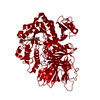
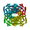
 Structure visualization
 Downloads & links
 Other downloads
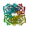


 PDBj
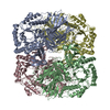
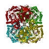
PDBj Assembly
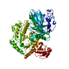


 Mass: 92.094 Da / Num. of mol.: 2 / Source method: obtained synthetically / Formula: C3H8O3
 Mass: 62.068 Da / Num. of mol.: 1 / Source method: obtained synthetically / Formula: C2H6O2
 Mass: 94.971 Da / Num. of mol.: 1 / Source method: obtained synthetically / Formula: PO4
 Mass: 24.305 Da / Num. of mol.: 2 / Source method: obtained synthetically / Formula: Mg
 Sample preparation
 / Beamline: ID14-1 / Wavelength: 0.933
  Processing