+ Open data
- Basic information
| Entry | Database: PDB / ID: 6wuc | |||||||||||||||||||||||||||||||||||||||||||||
|---|---|---|---|---|---|---|---|---|---|---|---|---|---|---|---|---|---|---|---|---|---|---|---|---|---|---|---|---|---|---|---|---|---|---|---|---|---|---|---|---|---|---|---|---|---|---|
| Title | The yeast Ctf3 complex with Cnn1-Wip1 | |||||||||||||||||||||||||||||||||||||||||||||
 Components Components |
| |||||||||||||||||||||||||||||||||||||||||||||
 Keywords Keywords | CELL CYCLE / kinetochore / chromosome segregation / CCAN | |||||||||||||||||||||||||||||||||||||||||||||
| Function / homology |  Function and homology information Function and homology informationnegative regulation of kinetochore assembly / attachment of spindle microtubules to kinetochore / centromeric DNA binding / establishment of mitotic sister chromatid cohesion / mitotic spindle assembly checkpoint signaling / DNA replication initiation / meiotic cell cycle / chromosome segregation / kinetochore / cell division ...negative regulation of kinetochore assembly / attachment of spindle microtubules to kinetochore / centromeric DNA binding / establishment of mitotic sister chromatid cohesion / mitotic spindle assembly checkpoint signaling / DNA replication initiation / meiotic cell cycle / chromosome segregation / kinetochore / cell division / protein-containing complex binding / nucleus Similarity search - Function | |||||||||||||||||||||||||||||||||||||||||||||
| Biological species |  | |||||||||||||||||||||||||||||||||||||||||||||
| Method | ELECTRON MICROSCOPY / single particle reconstruction / cryo EM / Resolution: 3.23 Å | |||||||||||||||||||||||||||||||||||||||||||||
 Authors Authors | Hinshaw, S.M. / Harrison, S.C. | |||||||||||||||||||||||||||||||||||||||||||||
| Funding support |  United States, 1items United States, 1items
| |||||||||||||||||||||||||||||||||||||||||||||
 Citation Citation |  Journal: Curr Biol / Year: 2020 Journal: Curr Biol / Year: 2020Title: The Structural Basis for Kinetochore Stabilization by Cnn1/CENP-T. Authors: Stephen M Hinshaw / Stephen C Harrison /  Abstract: Chromosome segregation depends on a regulated connection between spindle microtubules and centromeric DNA. The kinetochore mediates this connection and ensures it persists during anaphase, when ...Chromosome segregation depends on a regulated connection between spindle microtubules and centromeric DNA. The kinetochore mediates this connection and ensures it persists during anaphase, when sister chromatids must transit into daughter cells uninterrupted. The Ctf19 complex (Ctf19c) forms the centromeric base of the kinetochore in budding yeast. Biochemical experiments show that Ctf19c members associate hierarchically when purified from cell extract [1], an observation that is mostly explained by the structure of the complex [2]. The Ctf3 complex (Ctf3c), which is not required for the assembly of most other Ctf19c factors, disobeys the biochemical assembly hierarchy when observed in dividing cells that lack more basal components [3]. Thus, the biochemical experiments do not completely recapitulate the logic of centromeric Ctf19c assembly. We now present a high-resolution structure of the Ctf3c bound to the Cnn1-Wip1 heterodimer. Associated live-cell imaging experiments provide a mechanism for Ctf3c and Cnn1-Wip1 recruitment to the kinetochore. The mechanism suggests feedback regulation of Ctf19c assembly and unanticipated similarities in kinetochore organization between yeast and vertebrates. | |||||||||||||||||||||||||||||||||||||||||||||
| History |
|
- Structure visualization
| Movie |
 Movie viewer Movie viewer |
|---|---|
| Structure viewer | Molecule:  Molmil Molmil Jmol/JSmol Jmol/JSmol |
- Downloads & links
- Download
| PDBx/mmCIF format |  6wuc.cif.gz 6wuc.cif.gz | 248.7 KB | Display |  PDBx/mmCIF format PDBx/mmCIF format |
|---|---|---|---|---|
| PDB format |  pdb6wuc.ent.gz pdb6wuc.ent.gz | 193.7 KB | Display |  PDB format PDB format |
| PDBx/mmJSON format |  6wuc.json.gz 6wuc.json.gz | Tree view |  PDBx/mmJSON format PDBx/mmJSON format | |
| Others |  Other downloads Other downloads |
-Validation report
| Arichive directory |  https://data.pdbj.org/pub/pdb/validation_reports/wu/6wuc https://data.pdbj.org/pub/pdb/validation_reports/wu/6wuc ftp://data.pdbj.org/pub/pdb/validation_reports/wu/6wuc ftp://data.pdbj.org/pub/pdb/validation_reports/wu/6wuc | HTTPS FTP |
|---|
-Related structure data
| Related structure data |  21910MC M: map data used to model this data C: citing same article ( |
|---|---|
| Similar structure data |
- Links
Links
- Assembly
| Deposited unit | 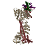
|
|---|---|
| 1 |
|
- Components
| #1: Protein | Mass: 21438.359 Da / Num. of mol.: 1 Source method: isolated from a genetically manipulated source Source: (gene. exp.)  Production host:  |
|---|---|
| #2: Protein | Mass: 84617.891 Da / Num. of mol.: 1 Source method: isolated from a genetically manipulated source Source: (gene. exp.)  Production host:  |
| #3: Protein | Mass: 27874.799 Da / Num. of mol.: 1 Source method: isolated from a genetically manipulated source Source: (gene. exp.)  Production host:  |
| #4: Protein | Mass: 10527.717 Da / Num. of mol.: 1 Source method: isolated from a genetically manipulated source Source: (gene. exp.)  Gene: WIP1 / Production host:  Trichoplusia ni (cabbage looper) / References: UniProt: Q2V2P8 Trichoplusia ni (cabbage looper) / References: UniProt: Q2V2P8 |
| #5: Protein | Mass: 41359.785 Da / Num. of mol.: 1 Source method: isolated from a genetically manipulated source Source: (gene. exp.)  Gene: CNN1 / Production host:  Trichoplusia ni (cabbage looper) / References: UniProt: P43618 Trichoplusia ni (cabbage looper) / References: UniProt: P43618 |
| Has protein modification | N |
-Experimental details
-Experiment
| Experiment | Method: ELECTRON MICROSCOPY |
|---|---|
| EM experiment | Aggregation state: PARTICLE / 3D reconstruction method: single particle reconstruction |
- Sample preparation
| Component | Name: Single particle reconstruction of the Ctf3c bound to Cnn1-Wip1 Type: COMPLEX / Entity ID: all / Source: RECOMBINANT |
|---|---|
| Source (natural) | Organism:  |
| Source (recombinant) | Organism:  |
| Buffer solution | pH: 8.5 |
| Specimen | Embedding applied: NO / Shadowing applied: NO / Staining applied: NO / Vitrification applied: YES |
| Vitrification | Cryogen name: ETHANE |
- Electron microscopy imaging
| Experimental equipment |  Model: Titan Krios / Image courtesy: FEI Company |
|---|---|
| Microscopy | Model: FEI TITAN KRIOS |
| Electron gun | Electron source:  FIELD EMISSION GUN / Accelerating voltage: 300 kV / Illumination mode: FLOOD BEAM FIELD EMISSION GUN / Accelerating voltage: 300 kV / Illumination mode: FLOOD BEAM |
| Electron lens | Mode: BRIGHT FIELD |
| Image recording | Electron dose: 60 e/Å2 / Film or detector model: GATAN K3 BIOQUANTUM (6k x 4k) |
- Processing
| Software | Name: PHENIX / Version: 1.17.1_3660: / Classification: refinement | ||||||||||||||||||||||||
|---|---|---|---|---|---|---|---|---|---|---|---|---|---|---|---|---|---|---|---|---|---|---|---|---|---|
| EM software |
| ||||||||||||||||||||||||
| CTF correction | Type: PHASE FLIPPING AND AMPLITUDE CORRECTION | ||||||||||||||||||||||||
| Symmetry | Point symmetry: C1 (asymmetric) | ||||||||||||||||||||||||
| 3D reconstruction | Resolution: 3.23 Å / Resolution method: FSC 0.143 CUT-OFF / Num. of particles: 109996 / Symmetry type: POINT | ||||||||||||||||||||||||
| Refine LS restraints |
|
 Movie
Movie Controller





 PDBj
PDBj