National Institutes of Health/National Institute of General Medical Sciences (NIH/NIGMS)
R35GM122510
米国
引用
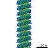
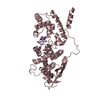
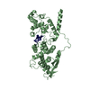
ジャーナル: Proc Natl Acad Sci U S A / 年: 2020 タイトル: Structures of filamentous viruses infecting hyperthermophilic archaea explain DNA stabilization in extreme environments. 著者: Fengbin Wang / Diana P Baquero / Leticia C Beltran / Zhangli Su / Tomasz Osinski / Weili Zheng / David Prangishvili / Mart Krupovic / Edward H Egelman / 要旨: Living organisms expend metabolic energy to repair and maintain their genomes, while viruses protect their genetic material by completely passive means. We have used cryo-electron microscopy (cryo-EM) ...Living organisms expend metabolic energy to repair and maintain their genomes, while viruses protect their genetic material by completely passive means. We have used cryo-electron microscopy (cryo-EM) to solve the atomic structures of two filamentous double-stranded DNA viruses that infect archaeal hosts living in nearly boiling acid: rod-shaped virus 1 (SSRV1), at 2.8-Å resolution, and filamentous virus (SIFV), at 4.0-Å resolution. The SIFV nucleocapsid is formed by a heterodimer of two homologous proteins and is membrane enveloped, while SSRV1 has a nucleocapsid formed by a homodimer and is not enveloped. In both, the capsid proteins wrap around the DNA and maintain it in an A-form. We suggest that the A-form is due to both a nonspecific desolvation of the DNA by the protein, and a specific coordination of the DNA phosphate groups by positively charged residues. We extend these observations by comparisons with four other archaeal filamentous viruses whose structures we have previously determined, and show that all 10 capsid proteins (from four heterodimers and two homodimers) have obvious structural homology while sequence similarity can be nonexistent. This arises from most capsid residues not being under any strong selective pressure. The inability to detect homology at the sequence level arises from the sampling of viruses in this part of the biosphere being extremely sparse. Comparative structural and genomic analyses suggest that nonenveloped archaeal viruses have evolved from enveloped viruses by shedding the membrane, indicating that this trait may be relatively easily lost during virus evolution.
履歴
登録
2020年4月28日
登録サイト: RCSB / 処理サイト: RCSB
改定 1.0
2020年7月29日
Provider: repository / タイプ: Initial release
改定 1.0
2020年7月29日
Data content type: EM metadata / Data content type: EM metadata / Provider: repository / タイプ: Initial release
改定 1.0
2020年7月29日
Data content type: Image / Data content type: Image / Provider: repository / タイプ: Initial release
改定 1.0
2020年7月29日
Data content type: Primary map / Data content type: Primary map / Provider: repository / タイプ: Initial release
改定 1.0
2020年7月29日
Data content type: Image / Data content type: Image / Provider: repository / タイプ: Initial release
改定 1.0
2020年7月29日
Data content type: Primary map / Data content type: Primary map / Provider: repository / タイプ: Initial release
改定 1.0
2020年7月29日
Data content type: Image / Data content type: Image / Provider: repository / タイプ: Initial release
改定 1.0
2020年7月29日
Data content type: Primary map / Data content type: Primary map / Provider: repository / タイプ: Initial release
Data content type: EM metadata / Data content type: EM metadata / EM metadata / Group: Data processing / Experimental summary / Data content type: EM metadata / EM metadata / カテゴリ: em_admin / em_software / Data content type: EM metadata / EM metadata / Item: _em_admin.last_update / _em_software.name
#241 - 2020年1月 20年の分子を振り返って (Twenty Years of Molecules) 類似性 (2)
-
集合体
登録構造単位
7: DNA (301-MER) 8: DNA (301-MER) A: Structural protein B: Structural protein C: Structural protein D: Structural protein E: Structural protein F: Structural protein G: Structural protein H: Structural protein I: Structural protein J: Structural protein K: Structural protein L: Structural protein M: Structural protein N: Structural protein O: Structural protein P: Structural protein Q: Structural protein R: Structural protein S: Structural protein T: Structural protein U: Structural protein V: Structural protein W: Structural protein a: Structural protein b: Structural protein c: Structural protein d: Structural protein e: Structural protein f: Structural protein g: Structural protein h: Structural protein i: Structural protein j: Structural protein k: Structural protein l: Structural protein m: Structural protein n: Structural protein o: Structural protein p: Structural protein q: Structural protein r: Structural protein s: Structural protein t: Structural protein u: Structural protein v: Structural protein w: Structural protein
根拠: microscopy, helical filament was observed by negative staining and Cryo-EM
タイプ
名称
対称操作
数
identity operation
1_555
1
Buried area
370700 Å2
ΔGint
-2578 kcal/mol
Surface area
185600 Å2
-
要素
#1: DNA鎖
DNA (301-MER)
分子量: 92878.148 Da / 分子数: 1 / 由来タイプ: 天然 / 由来: (天然) unclassified Rudivirus (ウイルス) Plasmid details: It is rod-like virus purified from a S. solfataricus strain
 ムービー
ムービー コントローラー


 データを開く
 基本情報
基本情報 要素
要素 キーワード
キーワード 機能・相同性情報
 unclassified Rudivirus (ウイルス)
 データ登録者
 米国, 1件
  引用
引用
 構造の表示
構造の表示 ダウンロードとリンク
 その他のダウンロード


 PDBj
PDBj


 集合体
集合体
 試料調製
試料調製 電子顕微鏡撮影
 FIELD EMISSION GUN / 加速電圧: 300 kV / 照射モード: FLOOD BEAM
 解析
解析