 Movie
Movie Controller


+ Open data

- Basic information
| Entry | Database: EMDB / ID: EMD-21867 | |||||||||
|---|---|---|---|---|---|---|---|---|---|---|
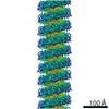
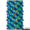
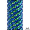
| Title | Cryo-EM of the S. solfataricus rod-shaped virus, SSRV1 | |||||||||
 Map data Map data | Cryo-EM of the S. solfataricus rod-shaped virus, SSRV1 | |||||||||
 Sample Sample | S. solfataricus rod-shaped virus, SSRV1 != unclassified Rudivirus S. solfataricus rod-shaped virus, SSRV1
| |||||||||
 Keywords Keywords | helical symmetry / archaeal virus / rod-like virus / structural protein / VIRUS | |||||||||
| Biological species |  unclassified Rudivirus unclassified Rudivirus | |||||||||
| Method | helical reconstruction / cryo EM / Resolution: 2.8 Å | |||||||||
 Authors Authors | Wang F / Baquero DP | |||||||||
| Funding support |  United States, 1 items United States, 1 items
| |||||||||
 Citation Citation | Journal: Proc Natl Acad Sci U S A / Year: 2020 Title: Structures of filamentous viruses infecting hyperthermophilic archaea explain DNA stabilization in extreme environments. Authors: Fengbin Wang / Diana P Baquero / Leticia C Beltran / Zhangli Su / Tomasz Osinski / Weili Zheng / David Prangishvili / Mart Krupovic / Edward H Egelman /  Abstract: Living organisms expend metabolic energy to repair and maintain their genomes, while viruses protect their genetic material by completely passive means. We have used cryo-electron microscopy (cryo-EM) ...Living organisms expend metabolic energy to repair and maintain their genomes, while viruses protect their genetic material by completely passive means. We have used cryo-electron microscopy (cryo-EM) to solve the atomic structures of two filamentous double-stranded DNA viruses that infect archaeal hosts living in nearly boiling acid: rod-shaped virus 1 (SSRV1), at 2.8-Å resolution, and filamentous virus (SIFV), at 4.0-Å resolution. The SIFV nucleocapsid is formed by a heterodimer of two homologous proteins and is membrane enveloped, while SSRV1 has a nucleocapsid formed by a homodimer and is not enveloped. In both, the capsid proteins wrap around the DNA and maintain it in an A-form. We suggest that the A-form is due to both a nonspecific desolvation of the DNA by the protein, and a specific coordination of the DNA phosphate groups by positively charged residues. We extend these observations by comparisons with four other archaeal filamentous viruses whose structures we have previously determined, and show that all 10 capsid proteins (from four heterodimers and two homodimers) have obvious structural homology while sequence similarity can be nonexistent. This arises from most capsid residues not being under any strong selective pressure. The inability to detect homology at the sequence level arises from the sampling of viruses in this part of the biosphere being extremely sparse. Comparative structural and genomic analyses suggest that nonenveloped archaeal viruses have evolved from enveloped viruses by shedding the membrane, indicating that this trait may be relatively easily lost during virus evolution. | |||||||||
| History |
|
- Structure visualization
| Movie |
 Movie viewer Movie viewer |
|---|---|
| Structure viewer | EM map: SurfViewMolmilJmol/JSmol |
| Supplemental images |

- Downloads & links
-EMDB archive
| Map data | emd_21867.map.gz | 78.7 MB | EMDB map data format | |
|---|---|---|---|---|
| Header (meta data) | emd-21867-v30.xmlemd-21867.xml | 16.3 KB 16.3 KB | Display Display | EMDB header |
| Images | 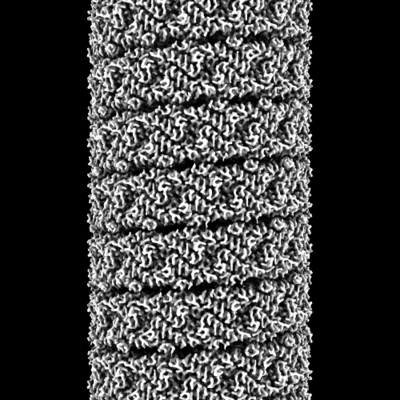 emd_21867.png emd_21867.png | 89.4 KB | ||
| Filedesc metadata | emd-21867.cif.gz | 5.7 KB | ||
| Archive directory |  http://ftp.pdbj.org/pub/emdb/structures/EMD-21867ftp://ftp.pdbj.org/pub/emdb/structures/EMD-21867 http://ftp.pdbj.org/pub/emdb/structures/EMD-21867ftp://ftp.pdbj.org/pub/emdb/structures/EMD-21867 | HTTPS FTP |
-Related structure data
| Related structure data | 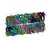 6wq0MC  6wq2C C: citing same article ( M: atomic model generated by this map |
|---|---|
| Similar structure data |


-Links
| EMDB pages | EMDB (EBI/PDBe) / EMDataResource |
|---|
-Map
| File | Download / File: emd_21867.map.gz / Format: CCP4 / Size: 125 MB / Type: IMAGE STORED AS FLOATING POINT NUMBER (4 BYTES) | ||||||||||||||||||||||||||||||||||||||||||||||||||||||||||||||||||||
|---|---|---|---|---|---|---|---|---|---|---|---|---|---|---|---|---|---|---|---|---|---|---|---|---|---|---|---|---|---|---|---|---|---|---|---|---|---|---|---|---|---|---|---|---|---|---|---|---|---|---|---|---|---|---|---|---|---|---|---|---|---|---|---|---|---|---|---|---|---|
| Annotation | Cryo-EM of the S. solfataricus rod-shaped virus, SSRV1 | ||||||||||||||||||||||||||||||||||||||||||||||||||||||||||||||||||||
| Projections & slices | Image control
Images are generated by Spider. | ||||||||||||||||||||||||||||||||||||||||||||||||||||||||||||||||||||
| Voxel size | X=Y=Z: 1.08 Å | ||||||||||||||||||||||||||||||||||||||||||||||||||||||||||||||||||||
| Density |
| ||||||||||||||||||||||||||||||||||||||||||||||||||||||||||||||||||||
| Symmetry | Space group: 1 | ||||||||||||||||||||||||||||||||||||||||||||||||||||||||||||||||||||
| Details | EMDB XML:
CCP4 map header:
| ||||||||||||||||||||||||||||||||||||||||||||||||||||||||||||||||||||
 Z (Sec.)
 Y (Row.)
 X (Col.)


-Supplemental data
- Sample components
-Entire : S. solfataricus rod-shaped virus, SSRV1
| Entire | Name: S. solfataricus rod-shaped virus, SSRV1 |
|---|---|
| Components |
|
-Supramolecule #1: unclassified Rudivirus
| Supramolecule | Name: unclassified Rudivirus / type: virus / ID: 1 / Parent: 0 / Macromolecule list: all / NCBI-ID: 351054 / Sci species name: unclassified Rudivirus / Virus type: VIRION / Virus isolate: STRAIN / Virus enveloped: No / Virus empty: No |
|---|---|
| Host (natural) | Organism:   Saccharolobus solfataricus (archaea) Saccharolobus solfataricus (archaea) |
-Macromolecule #1: DNA (301-MER)
| Macromolecule | Name: DNA (301-MER) / type: dna / ID: 1 / Number of copies: 1 / Classification: DNA |
|---|---|
| Source (natural) | Organism: unclassified Rudivirus |
| Molecular weight | Theoretical: 92.878148 KDa |
| Sequence | String: (DA)(DT)(DA)(DT)(DA)(DT)(DA)(DT)(DA)(DT) (DA)(DT)(DA)(DT)(DA)(DT)(DA)(DT)(DA)(DT) (DA)(DT)(DA)(DT)(DA)(DT)(DA)(DT)(DA) (DT)(DA)(DT)(DA)(DT)(DA)(DT)(DA)(DT)(DA) (DT) (DA)(DT)(DA)(DT)(DA)(DT) ...String: (DA)(DT)(DA)(DT)(DA)(DT)(DA)(DT)(DA)(DT) (DA)(DT)(DA)(DT)(DA)(DT)(DA)(DT)(DA)(DT) (DA)(DT)(DA)(DT)(DA)(DT)(DA)(DT)(DA) (DT)(DA)(DT)(DA)(DT)(DA)(DT)(DA)(DT)(DA) (DT) (DA)(DT)(DA)(DT)(DA)(DT)(DA)(DT) (DA)(DT)(DA)(DT)(DA)(DT)(DA)(DT)(DA)(DT) (DA)(DT) (DA)(DT)(DA)(DT)(DA)(DT)(DA) (DT)(DA)(DT)(DA)(DT)(DA)(DT)(DA)(DT)(DA) (DT)(DA)(DT) (DA)(DT)(DA)(DT)(DA)(DT) (DA)(DT)(DA)(DT)(DA)(DT)(DA)(DT)(DA)(DT) (DA)(DT)(DA)(DT) (DA)(DT)(DA)(DT)(DA) (DT)(DA)(DT)(DA)(DT)(DA)(DT)(DA)(DT)(DA) (DT)(DA)(DT)(DA)(DT) (DA)(DT)(DA)(DT) (DA)(DT)(DA)(DT)(DA)(DT)(DA)(DT)(DA)(DT) (DA)(DT)(DA)(DT)(DA)(DT) (DA)(DT)(DA) (DT)(DA)(DT)(DA)(DT)(DA)(DT)(DA)(DT)(DA) (DT)(DA)(DT)(DA)(DT)(DA)(DT) (DA)(DT) (DA)(DT)(DA)(DT)(DA)(DT)(DA)(DT)(DA)(DT) (DA)(DT)(DA)(DT)(DA)(DT)(DA)(DT) (DA) (DT)(DA)(DT)(DA)(DT)(DA)(DT)(DA)(DT)(DA) (DT)(DA)(DT)(DA)(DT)(DA)(DT)(DA)(DT) (DA)(DT)(DA)(DT)(DA)(DT)(DA)(DT)(DA)(DT) (DA)(DT)(DA)(DT)(DA)(DT)(DA)(DT)(DA)(DT) (DA)(DT)(DA)(DT)(DA)(DT)(DA)(DT)(DA) (DT)(DA)(DT)(DA)(DT)(DA)(DT)(DA)(DT)(DA) (DT) (DA)(DT)(DA)(DT)(DA)(DT)(DA)(DT) (DA)(DT)(DA)(DT)(DA)(DT)(DA)(DT)(DA)(DT) (DA)(DT) (DA)(DT)(DA)(DT)(DA)(DT)(DA) (DT)(DA)(DT)(DA)(DT)(DA)(DT)(DA)(DT)(DA) (DT)(DA)(DT) (DA)(DT)(DA)(DT)(DA)(DT) (DA)(DT)(DA)(DT)(DA)(DT)(DA)(DT)(DA)(DT) (DA)(DT)(DA)(DT) (DA) |
-Macromolecule #2: DNA (301-MER)
| Macromolecule | Name: DNA (301-MER) / type: dna / ID: 2 / Number of copies: 1 / Classification: DNA |
|---|---|
| Source (natural) | Organism: unclassified Rudivirus |
| Molecular weight | Theoretical: 92.869141 KDa |
| Sequence | String: (DT)(DA)(DT)(DA)(DT)(DA)(DT)(DA)(DT)(DA) (DT)(DA)(DT)(DA)(DT)(DA)(DT)(DA)(DT)(DA) (DT)(DA)(DT)(DA)(DT)(DA)(DT)(DA)(DT) (DA)(DT)(DA)(DT)(DA)(DT)(DA)(DT)(DA)(DT) (DA) (DT)(DA)(DT)(DA)(DT)(DA) ...String: (DT)(DA)(DT)(DA)(DT)(DA)(DT)(DA)(DT)(DA) (DT)(DA)(DT)(DA)(DT)(DA)(DT)(DA)(DT)(DA) (DT)(DA)(DT)(DA)(DT)(DA)(DT)(DA)(DT) (DA)(DT)(DA)(DT)(DA)(DT)(DA)(DT)(DA)(DT) (DA) (DT)(DA)(DT)(DA)(DT)(DA)(DT)(DA) (DT)(DA)(DT)(DA)(DT)(DA)(DT)(DA)(DT)(DA) (DT)(DA) (DT)(DA)(DT)(DA)(DT)(DA)(DT) (DA)(DT)(DA)(DT)(DA)(DT)(DA)(DT)(DA)(DT) (DA)(DT)(DA) (DT)(DA)(DT)(DA)(DT)(DA) (DT)(DA)(DT)(DA)(DT)(DA)(DT)(DA)(DT)(DA) (DT)(DA)(DT)(DA) (DT)(DA)(DT)(DA)(DT) (DA)(DT)(DA)(DT)(DA)(DT)(DA)(DT)(DA)(DT) (DA)(DT)(DA)(DT)(DA) (DT)(DA)(DT)(DA) (DT)(DA)(DT)(DA)(DT)(DA)(DT)(DA)(DT)(DA) (DT)(DA)(DT)(DA)(DT)(DA) (DT)(DA)(DT) (DA)(DT)(DA)(DT)(DA)(DT)(DA)(DT)(DA)(DT) (DA)(DT)(DA)(DT)(DA)(DT)(DA) (DT)(DA) (DT)(DA)(DT)(DA)(DT)(DA)(DT)(DA)(DT)(DA) (DT)(DA)(DT)(DA)(DT)(DA)(DT)(DA) (DT) (DA)(DT)(DA)(DT)(DA)(DT)(DA)(DT)(DA)(DT) (DA)(DT)(DA)(DT)(DA)(DT)(DA)(DT)(DA) (DT)(DA)(DT)(DA)(DT)(DA)(DT)(DA)(DT)(DA) (DT)(DA)(DT)(DA)(DT)(DA)(DT)(DA)(DT)(DA) (DT)(DA)(DT)(DA)(DT)(DA)(DT)(DA)(DT) (DA)(DT)(DA)(DT)(DA)(DT)(DA)(DT)(DA)(DT) (DA) (DT)(DA)(DT)(DA)(DT)(DA)(DT)(DA) (DT)(DA)(DT)(DA)(DT)(DA)(DT)(DA)(DT)(DA) (DT)(DA) (DT)(DA)(DT)(DA)(DT)(DA)(DT) (DA)(DT)(DA)(DT)(DA)(DT)(DA)(DT)(DA)(DT) (DA)(DT)(DA) (DT)(DA)(DT)(DA)(DT)(DA) (DT)(DA)(DT)(DA)(DT)(DA)(DT)(DA)(DT)(DA) (DT)(DA)(DT)(DA) (DT) |
-Macromolecule #3: Structural protein
| Macromolecule | Name: Structural protein / type: protein_or_peptide / ID: 3 / Number of copies: 46 / Enantiomer: LEVO |
|---|---|
| Source (natural) | Organism: unclassified Rudivirus |
| Molecular weight | Theoretical: 14.207113 KDa |
| Sequence | String: MAKGRTPRSF SQRYGKWNAK FTAFSNPTVA STILTNVAPI AQGNFQTNVP KFTSVNEQVS AVLTQYGVTG PSRAIYQGYG LKVARALNR IGAGPALTNM VAGLKAYYVS AYGANPEILD AVTNIILGSP TGYVS |
-Experimental details
-Structure determination
| Method | cryo EM |
|---|---|
 Processing Processing | helical reconstruction |
| Aggregation state | filament |
-Sample preparation
| Buffer | pH: 6 |
|---|---|
| Grid | Details: unspecified |
| Vitrification | Cryogen name: ETHANE |
- Electron microscopy
| Microscope | FEI TITAN KRIOS |
|---|---|
| Image recording | Film or detector model: GATAN K3 (6k x 4k) / Average electron dose: 50.0 e/Å2 |
| Electron beam | Acceleration voltage: 300 kV / Electron source:  FIELD EMISSION GUN FIELD EMISSION GUN |
| Electron optics | Illumination mode: FLOOD BEAM / Imaging mode: BRIGHT FIELD |
| Experimental equipment |  Model: Titan Krios / Image courtesy: FEI Company |
-Image processing
| Final reconstruction | Applied symmetry - Helical parameters - Δz: 2.94 Å Applied symmetry - Helical parameters - Δ&Phi: 24.53 ° Applied symmetry - Helical parameters - Axial symmetry: D1 (2x1 fold dihedral) Resolution.type: BY AUTHOR / Resolution: 2.8 Å / Resolution method: OTHER / Details: MODEL:MAP FSC, D99, MAP:MAP FSC / Number images used: 470000 |
|---|---|
| CTF correction | Type: PHASE FLIPPING AND AMPLITUDE CORRECTION |
| Startup model | Type of model: OTHER Details: averaged cylinder using all segments, with random azimuthal angles |
| Final angle assignment | Type: NOT APPLICABLE |
