National Institutes of Health/National Institute of General Medical Sciences (NIH/NIGMS)
R35GM122510
United States
Citation
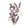
Journal: Proc Natl Acad Sci U S A / Year: 2020 Title: Structures of filamentous viruses infecting hyperthermophilic archaea explain DNA stabilization in extreme environments. Authors: Fengbin Wang / Diana P Baquero / Leticia C Beltran / Zhangli Su / Tomasz Osinski / Weili Zheng / David Prangishvili / Mart Krupovic / Edward H Egelman / Abstract: Living organisms expend metabolic energy to repair and maintain their genomes, while viruses protect their genetic material by completely passive means. We have used cryo-electron microscopy (cryo-EM) ...Living organisms expend metabolic energy to repair and maintain their genomes, while viruses protect their genetic material by completely passive means. We have used cryo-electron microscopy (cryo-EM) to solve the atomic structures of two filamentous double-stranded DNA viruses that infect archaeal hosts living in nearly boiling acid: rod-shaped virus 1 (SSRV1), at 2.8-Å resolution, and filamentous virus (SIFV), at 4.0-Å resolution. The SIFV nucleocapsid is formed by a heterodimer of two homologous proteins and is membrane enveloped, while SSRV1 has a nucleocapsid formed by a homodimer and is not enveloped. In both, the capsid proteins wrap around the DNA and maintain it in an A-form. We suggest that the A-form is due to both a nonspecific desolvation of the DNA by the protein, and a specific coordination of the DNA phosphate groups by positively charged residues. We extend these observations by comparisons with four other archaeal filamentous viruses whose structures we have previously determined, and show that all 10 capsid proteins (from four heterodimers and two homodimers) have obvious structural homology while sequence similarity can be nonexistent. This arises from most capsid residues not being under any strong selective pressure. The inability to detect homology at the sequence level arises from the sampling of viruses in this part of the biosphere being extremely sparse. Comparative structural and genomic analyses suggest that nonenveloped archaeal viruses have evolved from enveloped viruses by shedding the membrane, indicating that this trait may be relatively easily lost during virus evolution.
History
Deposition
Apr 28, 2020
Deposition site: RCSB / Processing site: RCSB
Revision 1.0
Jul 29, 2020
Provider: repository / Type: Initial release
Revision 1.0
Jul 29, 2020
Data content type: EM metadata / Data content type: EM metadata / Provider: repository / Type: Initial release
Revision 1.0
Jul 29, 2020
Data content type: Image / Data content type: Image / Provider: repository / Type: Initial release
Revision 1.0
Jul 29, 2020
Data content type: Primary map / Data content type: Primary map / Provider: repository / Type: Initial release
Revision 1.0
Jul 29, 2020
Data content type: Image / Data content type: Image / Provider: repository / Type: Initial release
Revision 1.0
Jul 29, 2020
Data content type: Primary map / Data content type: Primary map / Provider: repository / Type: Initial release
Revision 1.0
Jul 29, 2020
Data content type: Image / Data content type: Image / Provider: repository / Type: Initial release
Revision 1.0
Jul 29, 2020
Data content type: Primary map / Data content type: Primary map / Provider: repository / Type: Initial release
Data content type: EM metadata / Data content type: EM metadata / EM metadata / Group: Data processing / Experimental summary / Data content type: EM metadata / EM metadata / Category: em_admin / em_software / Data content type: EM metadata / EM metadata / Item: _em_admin.last_update / _em_software.name
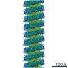
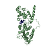
7: DNA (301-MER) 8: DNA (301-MER) A: Structural protein B: Structural protein C: Structural protein D: Structural protein E: Structural protein F: Structural protein G: Structural protein H: Structural protein I: Structural protein J: Structural protein K: Structural protein L: Structural protein M: Structural protein N: Structural protein O: Structural protein P: Structural protein Q: Structural protein R: Structural protein S: Structural protein T: Structural protein U: Structural protein V: Structural protein W: Structural protein a: Structural protein b: Structural protein c: Structural protein d: Structural protein e: Structural protein f: Structural protein g: Structural protein h: Structural protein i: Structural protein j: Structural protein k: Structural protein l: Structural protein m: Structural protein n: Structural protein o: Structural protein p: Structural protein q: Structural protein r: Structural protein s: Structural protein t: Structural protein u: Structural protein v: Structural protein w: Structural protein
Evidence: microscopy, helical filament was observed by negative staining and Cryo-EM
Type
Name
Symmetry operation
Number
identity operation
1_555
1
Buried area
370700 Å2
ΔGint
-2578 kcal/mol
Surface area
185600 Å2
-
Components
#1: DNA chain
DNA (301-MER)
Mass: 92878.148 Da / Num. of mol.: 1 / Source method: isolated from a natural source / Source: (natural) unclassified Rudivirus Plasmid details: It is rod-like virus purified from a S. solfataricus strain
#2: DNA chain
DNA (301-MER)
Mass: 92869.141 Da / Num. of mol.: 1 / Source method: isolated from a natural source / Source: (natural) unclassified Rudivirus
#3: Protein
... Structuralprotein
Mass: 14207.113 Da / Num. of mol.: 46 / Source method: isolated from a natural source / Source: (natural) unclassified Rudivirus
Has protein modification
N
-
Experimental details
-
Experiment
Experiment
Method: ELECTRON MICROSCOPY
EM experiment
Aggregation state: FILAMENT / 3D reconstruction method: helical reconstruction
-
Sample preparation
Component
Name: S. solfataricus rod-shaped virus, SSRV1 / Type: VIRUS / Entity ID: all / Source: NATURAL
Source (natural)
Organism: unclassified Rudivirus
Details of virus
Empty: NO / Enveloped: NO / Isolate: STRAIN / Type: VIRION
Natural host
Organism: Saccharolobus solfataricus
Buffer solution
pH: 6
Specimen
Embedding applied: NO / Shadowing applied: NO / Staining applied: NO / Vitrification applied: YES
Specimen support
Details: unspecified
Vitrification
Cryogen name: ETHANE
-
Electron microscopy imaging
Experimental equipment
Model: Titan Krios / Image courtesy: FEI Company
Microscopy
Model: FEI TITAN KRIOS
Electron gun
Electron source: FIELD EMISSION GUN / Accelerating voltage: 300 kV / Illumination mode: FLOOD BEAM
Electron lens
Mode: BRIGHT FIELD
Image recording
Electron dose: 50 e/Å2 / Film or detector model: GATAN K3 (6k x 4k)
In the structure databanks used in Yorodumi, some data are registered as the other names, "COVID-19 virus" and "2019-nCoV". Here are the details of the virus and the list of structure data.
Jan 31, 2019. EMDB accession codes are about to change! (news from PDBe EMDB page)
EMDB accession codes are about to change! (news from PDBe EMDB page)
The allocation of 4 digits for EMDB accession codes will soon come to an end. Whilst these codes will remain in use, new EMDB accession codes will include an additional digit and will expand incrementally as the available range of codes is exhausted. The current 4-digit format prefixed with “EMD-” (i.e. EMD-XXXX) will advance to a 5-digit format (i.e. EMD-XXXXX), and so on. It is currently estimated that the 4-digit codes will be depleted around Spring 2019, at which point the 5-digit format will come into force.
The EM Navigator/Yorodumi systems omit the EMD- prefix.
Related info.:Q: What is EMD? / ID/Accession-code notation in Yorodumi/EM Navigator
Yorodumi is a browser for structure data from EMDB, PDB, SASBDB, etc.
This page is also the successor to EM Navigator detail page, and also detail information page/front-end page for Omokage search.
The word "yorodu" (or yorozu) is an old Japanese word meaning "ten thousand". "mi" (miru) is to see.
Related info.:EMDB / PDB / SASBDB / Comparison of 3 databanks / Yorodumi Search / Aug 31, 2016. New EM Navigator & Yorodumi / Yorodumi Papers / Jmol/JSmol / Function and homology information / Changes in new EM Navigator and Yorodumi
 Movie
Movie Controller


 Open data
 Basic information
 Components
 Keywords
 Function and homology information
 unclassified Rudivirus
 Authors
 United States, 1items
  Citation
 Structure visualization
 Downloads & links
 Other downloads


 PDBj
PDBj


 Assembly
 Sample preparation
 Electron microscopy imaging
 FIELD EMISSION GUN / Accelerating voltage: 300 kV / Illumination mode: FLOOD BEAM
 Processing