Movie
Movie Controller

[English] 日本語
 Yorodumi
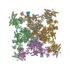
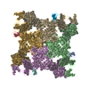
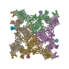
- PDB-6wot: Cryo-EM structure of recombinant rabbit Ryanodine Receptor type 1... -
+ Open data

- Basic information
| Entry | Database: PDB / ID: 6wot | ||||||||||||||||||||||||
|---|---|---|---|---|---|---|---|---|---|---|---|---|---|---|---|---|---|---|---|---|---|---|---|---|---|
| Title | Cryo-EM structure of recombinant rabbit Ryanodine Receptor type 1 mutant R164C in complex with FKBP12.6 | ||||||||||||||||||||||||
 Components Components |
| ||||||||||||||||||||||||
 Keywords Keywords | TRANSPORT PROTEIN/ISOMERASE / Ryanodine receptor / Calcium channel / Mutation / NTDA mutation / RyR1 / malignant hyperthermia / central core disease / TRANSPORT PROTEIN-ISOMERASE complex | ||||||||||||||||||||||||
| Function / homology |  Function and homology information Function and homology informationATP-gated ion channel activity / negative regulation of calcium-mediated signaling / ryanodine-sensitive calcium-release channel activity / terminal cisterna / ryanodine receptor complex / release of sequestered calcium ion into cytosol by sarcoplasmic reticulum / negative regulation of release of sequestered calcium ion into cytosol / response to redox state / ossification involved in bone maturation / cellular response to caffeine ...ATP-gated ion channel activity / negative regulation of calcium-mediated signaling / ryanodine-sensitive calcium-release channel activity / terminal cisterna / ryanodine receptor complex / release of sequestered calcium ion into cytosol by sarcoplasmic reticulum / negative regulation of release of sequestered calcium ion into cytosol / response to redox state / ossification involved in bone maturation / cellular response to caffeine / negative regulation of heart rate / skin development / 'de novo' protein folding / FK506 binding / organelle membrane / smooth endoplasmic reticulum / outflow tract morphogenesis / intracellularly gated calcium channel activity / regulation of ryanodine-sensitive calcium-release channel activity / toxic substance binding / voltage-gated calcium channel activity / calcium channel inhibitor activity / skeletal muscle fiber development / regulation of release of sequestered calcium ion into cytosol by sarcoplasmic reticulum / release of sequestered calcium ion into cytosol / Ion homeostasis / regulation of cardiac muscle contraction by regulation of the release of sequestered calcium ion / muscle contraction / sarcoplasmic reticulum membrane / calcium channel complex / striated muscle contraction / cellular response to calcium ion / peptidylprolyl isomerase / sarcoplasmic reticulum / peptidyl-prolyl cis-trans isomerase activity / calcium-mediated signaling / calcium channel regulator activity / protein maturation / sarcolemma / intracellular calcium ion homeostasis / Stimuli-sensing channels / Z disc / calcium channel activity / calcium ion transmembrane transport / disordered domain specific binding / protein refolding / protein folding / protein homotetramerization / transmembrane transporter binding / calmodulin binding / signaling receptor binding / calcium ion binding / ATP binding / membrane / identical protein binding / cytoplasm Similarity search - Function | ||||||||||||||||||||||||
| Biological species |   Homo sapiens (human) Homo sapiens (human) | ||||||||||||||||||||||||
| Method | ELECTRON MICROSCOPY / single particle reconstruction / cryo EM / Resolution: 3.54 Å | ||||||||||||||||||||||||
 Authors Authors | Iyer, K.A. / Hu, Y. / Kurebayashi, N. / Murayama, T. / Samso, M. | ||||||||||||||||||||||||
| Funding support |  United States, 7items United States, 7items
| ||||||||||||||||||||||||
 Citation Citation | Journal: Sci Adv / Year: 2020 Title: Structural mechanism of two gain-of-function cardiac and skeletal RyR mutations at an equivalent site by cryo-EM. Authors: Kavita A Iyer / Yifan Hu / Ashok R Nayak / Nagomi Kurebayashi / Takashi Murayama / Montserrat Samsó /  Abstract: Mutations in ryanodine receptors (RyRs), intracellular Ca channels, are associated with deadly disorders. Despite abundant functional studies, the molecular mechanism of RyR malfunction remains ...Mutations in ryanodine receptors (RyRs), intracellular Ca channels, are associated with deadly disorders. Despite abundant functional studies, the molecular mechanism of RyR malfunction remains elusive. We studied two single-point mutations at an equivalent site in the skeletal (RyR1 R164C) and cardiac (RyR2 R176Q) isoforms using ryanodine binding, Ca imaging, and cryo-electron microscopy (cryo-EM) of the full-length protein. Loss of the positive charge had greater effect on the skeletal isoform, mediated via distortion of a salt bridge network, a molecular latch inducing rotation of a cytoplasmic domain, and partial progression to open-state traits of the large cytoplasmic assembly accompanied by alteration of the Ca binding site, which concur with the major "hyperactive" feature of the mutated channel. Our cryo-EM studies demonstrated the allosteric effect of a mutation situated ~85 Å away from the pore and identified an isoform-specific structural effect. | ||||||||||||||||||||||||
| History |
|
- Structure visualization
| Movie |
Movie viewer |
|---|---|
| Structure viewer | Molecule: MolmilJmol/JSmol |
- Downloads & links
-Download
| PDBx/mmCIF format | 6wot.cif.gz | 5.1 MB | Display | PDBx/mmCIF format |
|---|---|---|---|---|
| PDB format | pdb6wot.ent.gz | Display | PDB format | |
| PDBx/mmJSON format | 6wot.json.gz | Tree view | PDBx/mmJSON format | |
| Others |  Other downloads Other downloads |
-Validation report
| Arichive directory | https://data.pdbj.org/pub/pdb/validation_reports/wo/6wotftp://data.pdbj.org/pub/pdb/validation_reports/wo/6wot | HTTPS FTP |
|---|
-Related structure data
| Related structure data |  21860MC  6wouC  6wovC M: map data used to model this data C: citing same article ( |
|---|---|
| Similar structure data |


-Links
 PDBj
PDBj


- Assembly
| Deposited unit | 
|
|---|---|
| 1 |
|
-Components
| #1: Protein | Mass: 565737.500 Da / Num. of mol.: 4 / Mutation: R164C Source method: isolated from a genetically manipulated source Source: (gene. exp.) Homo sapiens (human) / References: UniProt: P11716#2: Protein | Mass: 11667.305 Da / Num. of mol.: 4 Source method: isolated from a genetically manipulated source Source: (gene. exp.) Homo sapiens (human) / Gene: FKBP1B, FKBP12.6, FKBP1L, FKBP9, OTK4Production host:  References: UniProt: P68106, peptidylprolyl isomerase #3: Chemical | ChemComp-ZN /   Mass: 65.409 Da / Num. of mol.: 4 / Source method: obtained synthetically / Formula: Zn Mass: 65.409 Da / Num. of mol.: 4 / Source method: obtained synthetically / Formula: ZnHas ligand of interest | N | |
|---|
-Experimental details
-Experiment
| Experiment | Method: ELECTRON MICROSCOPY |
|---|---|
| EM experiment | Aggregation state: PARTICLE / 3D reconstruction method: single particle reconstruction |
- Sample preparation
| Component |
| |||||||||||||||||||||||||
|---|---|---|---|---|---|---|---|---|---|---|---|---|---|---|---|---|---|---|---|---|---|---|---|---|---|---|
| Molecular weight | Value: 2.26 MDa / Experimental value: NO | |||||||||||||||||||||||||
| Source (natural) |
| |||||||||||||||||||||||||
| Source (recombinant) |
| |||||||||||||||||||||||||
| Buffer solution | pH: 7.4 | |||||||||||||||||||||||||
| Buffer component |
| |||||||||||||||||||||||||
| Specimen | Conc.: 5 mg/ml / Embedding applied: NO / Shadowing applied: NO / Staining applied: NO / Vitrification applied: YES | |||||||||||||||||||||||||
| Specimen support | Grid material: GOLD / Grid mesh size: 300 divisions/in. / Grid type: Quantifoil, UltrAuFoil, R1.2/1.3 | |||||||||||||||||||||||||
| Vitrification | Instrument: FEI VITROBOT MARK IV / Cryogen name: ETHANE / Humidity: 95 % / Chamber temperature: 277 K |
- Electron microscopy imaging
| Experimental equipment |  Model: Titan Krios / Image courtesy: FEI Company |
|---|---|
| Microscopy | Model: FEI TITAN KRIOS |
| Electron gun | Electron source:  FIELD EMISSION GUN / Accelerating voltage: 300 kV / Illumination mode: FLOOD BEAM FIELD EMISSION GUN / Accelerating voltage: 300 kV / Illumination mode: FLOOD BEAM |
| Electron lens | Mode: BRIGHT FIELD / Nominal magnification: 81000 X / Nominal defocus max: 2000 nm / Nominal defocus min: 1000 nm / Cs: 2.7 mm / C2 aperture diameter: 100 µm |
| Specimen holder | Cryogen: NITROGEN / Specimen holder model: FEI TITAN KRIOS AUTOGRID HOLDER |
| Image recording | Average exposure time: 3.49 sec. / Electron dose: 50 e/Å2 / Film or detector model: GATAN K3 (6k x 4k) / Num. of grids imaged: 1 / Num. of real images: 4923 |
| EM imaging optics | Energyfilter slit width: 20 eV |
- Processing
| Software |
| ||||||||||||||||||||||||||||||||
|---|---|---|---|---|---|---|---|---|---|---|---|---|---|---|---|---|---|---|---|---|---|---|---|---|---|---|---|---|---|---|---|---|---|
| EM software |
| ||||||||||||||||||||||||||||||||
| CTF correction | Type: NONE | ||||||||||||||||||||||||||||||||
| Particle selection | Num. of particles selected: 581283 | ||||||||||||||||||||||||||||||||
| Symmetry | Point symmetry: C4 (4 fold cyclic) | ||||||||||||||||||||||||||||||||
| 3D reconstruction | Resolution: 3.54 Å / Resolution method: FSC 0.143 CUT-OFF / Num. of particles: 385324 / Symmetry type: POINT | ||||||||||||||||||||||||||||||||
| Atomic model building | Protocol: RIGID BODY FIT / Space: REAL | ||||||||||||||||||||||||||||||||
| Atomic model building | PDB-ID: 5TB0 Accession code: 5TB0 / Source name: PDB / Type: experimental model | ||||||||||||||||||||||||||||||||
| Refinement | Cross valid method: NONE Stereochemistry target values: GeoStd + Monomer Library + CDL v1.2 | ||||||||||||||||||||||||||||||||
| Displacement parameters | Biso mean: 223.74 Å2 | ||||||||||||||||||||||||||||||||
| Refine LS restraints |
|