 Movie
Movie Controller

[English] 日本語
 Yorodumi
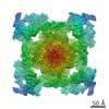
- PDB-6wov: Cryo-EM structure of recombinant mouse Ryanodine Receptor type 2 ... -
+ Open data

- Basic information
| Entry | Database: PDB / ID: 6wov | ||||||||||||||||||||||||
|---|---|---|---|---|---|---|---|---|---|---|---|---|---|---|---|---|---|---|---|---|---|---|---|---|---|
| Title | Cryo-EM structure of recombinant mouse Ryanodine Receptor type 2 wild type in complex with FKBP12.6 | ||||||||||||||||||||||||
 Components Components |
| ||||||||||||||||||||||||
 Keywords Keywords | TRANSPORT PROTEIN/ISOMERASE / Ryanodine receptor / Calcium channel / RyR2 / CICR / TRANSPORT PROTEIN-ISOMERASE complex / excitation-contraction coupling | ||||||||||||||||||||||||
| Function / homology |  Function and homology information Function and homology informationmanganese ion transmembrane transport / establishment of protein localization to endoplasmic reticulum / type B pancreatic cell apoptotic process / Purkinje myocyte to ventricular cardiac muscle cell signaling / regulation of atrial cardiac muscle cell action potential / left ventricular cardiac muscle tissue morphogenesis / suramin binding / regulation of AV node cell action potential / sarcoplasmic reticulum calcium ion transport / regulation of SA node cell action potential ...manganese ion transmembrane transport / establishment of protein localization to endoplasmic reticulum / type B pancreatic cell apoptotic process / Purkinje myocyte to ventricular cardiac muscle cell signaling / regulation of atrial cardiac muscle cell action potential / left ventricular cardiac muscle tissue morphogenesis / suramin binding / regulation of AV node cell action potential / sarcoplasmic reticulum calcium ion transport / regulation of SA node cell action potential / calcium-induced calcium release activity / A band / Stimuli-sensing channels / : / ventricular cardiac muscle cell action potential / negative regulation of calcium-mediated signaling / Ion homeostasis / regulation of ventricular cardiac muscle cell action potential / cardiac muscle hypertrophy / embryonic heart tube morphogenesis / negative regulation of insulin secretion involved in cellular response to glucose stimulus / neuronal action potential propagation / calcium ion transport into cytosol / insulin secretion involved in cellular response to glucose stimulus / ryanodine-sensitive calcium-release channel activity / release of sequestered calcium ion into cytosol by sarcoplasmic reticulum / negative regulation of release of sequestered calcium ion into cytosol / response to redox state / regulation of cardiac muscle contraction by calcium ion signaling / cellular response to caffeine / negative regulation of heart rate / extrinsic component of cytoplasmic side of plasma membrane / 'de novo' protein folding / response to caffeine / FK506 binding / response to muscle activity / calcium ion transmembrane import into cytosol / negative regulation of cytosolic calcium ion concentration / protein kinase A regulatory subunit binding / protein kinase A catalytic subunit binding / positive regulation of the force of heart contraction / intracellularly gated calcium channel activity / smooth endoplasmic reticulum / smooth muscle contraction / T cell proliferation / response to magnesium ion / detection of calcium ion / regulation of cardiac muscle contraction / regulation of cytosolic calcium ion concentration / positive regulation of heart rate / calcium channel inhibitor activity / striated muscle contraction / regulation of release of sequestered calcium ion into cytosol by sarcoplasmic reticulum / Ion homeostasis / response to muscle stretch / cardiac muscle contraction / release of sequestered calcium ion into cytosol / regulation of cardiac muscle contraction by regulation of the release of sequestered calcium ion / sarcoplasmic reticulum membrane / cellular response to epinephrine stimulus / calcium channel complex / regulation of heart rate / sarcoplasmic reticulum / peptidylprolyl isomerase / sarcomere / peptidyl-prolyl cis-trans isomerase activity / calcium channel regulator activity / calcium-mediated signaling / protein maturation / sarcolemma / response to calcium ion / Stimuli-sensing channels / Z disc / calcium ion transmembrane transport / calcium channel activity / intracellular calcium ion homeostasis / calcium ion transport / nuclear envelope / protein refolding / positive regulation of cytosolic calcium ion concentration / scaffold protein binding / monoatomic ion transmembrane transport / transmembrane transporter binding / response to hypoxia / calmodulin binding / signaling receptor binding / calcium ion binding / protein kinase binding / enzyme binding / protein-containing complex / membrane / identical protein binding / cytoplasm Similarity search - Function | ||||||||||||||||||||||||
| Biological species |   Homo sapiens (human) Homo sapiens (human) | ||||||||||||||||||||||||
| Method | ELECTRON MICROSCOPY / single particle reconstruction / cryo EM / Resolution: 5.1 Å | ||||||||||||||||||||||||
 Authors Authors | Iyer, K.A. / Hu, Y. / Nayak, A.R. / Kurebayashi, N. / Murayama, T. / Samso, M. | ||||||||||||||||||||||||
| Funding support |  United States, 7items United States, 7items
| ||||||||||||||||||||||||
 Citation Citation | Journal: Sci Adv / Year: 2020 Title: Structural mechanism of two gain-of-function cardiac and skeletal RyR mutations at an equivalent site by cryo-EM. Authors: Kavita A Iyer / Yifan Hu / Ashok R Nayak / Nagomi Kurebayashi / Takashi Murayama / Montserrat Samsó /  Abstract: Mutations in ryanodine receptors (RyRs), intracellular Ca channels, are associated with deadly disorders. Despite abundant functional studies, the molecular mechanism of RyR malfunction remains ...Mutations in ryanodine receptors (RyRs), intracellular Ca channels, are associated with deadly disorders. Despite abundant functional studies, the molecular mechanism of RyR malfunction remains elusive. We studied two single-point mutations at an equivalent site in the skeletal (RyR1 R164C) and cardiac (RyR2 R176Q) isoforms using ryanodine binding, Ca imaging, and cryo-electron microscopy (cryo-EM) of the full-length protein. Loss of the positive charge had greater effect on the skeletal isoform, mediated via distortion of a salt bridge network, a molecular latch inducing rotation of a cytoplasmic domain, and partial progression to open-state traits of the large cytoplasmic assembly accompanied by alteration of the Ca binding site, which concur with the major "hyperactive" feature of the mutated channel. Our cryo-EM studies demonstrated the allosteric effect of a mutation situated ~85 Å away from the pore and identified an isoform-specific structural effect. | ||||||||||||||||||||||||
| History |
|
- Structure visualization
| Movie |
Movie viewer |
|---|---|
| Structure viewer | Molecule: MolmilJmol/JSmol |
- Downloads & links
-Download
| PDBx/mmCIF format | 6wov.cif.gz | 5 MB | Display | PDBx/mmCIF format |
|---|---|---|---|---|
| PDB format | pdb6wov.ent.gz | Display | PDB format | |
| PDBx/mmJSON format | 6wov.json.gz | Tree view | PDBx/mmJSON format | |
| Others |  Other downloads Other downloads |
-Validation report
| Arichive directory | https://data.pdbj.org/pub/pdb/validation_reports/wo/6wovftp://data.pdbj.org/pub/pdb/validation_reports/wo/6wov | HTTPS FTP |
|---|
-Related structure data
| Related structure data |  21862MC  6wotC  6wouC M: map data used to model this data C: citing same article ( |
|---|---|
| Similar structure data |

-Links
 PDBj
PDBj


- Assembly
| Deposited unit | 
|
|---|---|
| 1 |
|
-Components
| #1: Protein | Mass: 565536.000 Da / Num. of mol.: 4 Source method: isolated from a genetically manipulated source Source: (gene. exp.) Homo sapiens (human) / References: UniProt: E9Q401#2: Protein | Mass: 11667.305 Da / Num. of mol.: 4 Source method: isolated from a genetically manipulated source Source: (gene. exp.) Homo sapiens (human) / Gene: FKBP1B, FKBP12.6, FKBP1L, FKBP9, OTK4Production host:  References: UniProt: P68106, peptidylprolyl isomerase #3: Chemical | ChemComp-ZN /   Mass: 65.409 Da / Num. of mol.: 4 / Source method: obtained synthetically / Formula: Zn Mass: 65.409 Da / Num. of mol.: 4 / Source method: obtained synthetically / Formula: ZnHas ligand of interest | N | |
|---|
-Experimental details
-Experiment
| Experiment | Method: ELECTRON MICROSCOPY |
|---|---|
| EM experiment | Aggregation state: PARTICLE / 3D reconstruction method: single particle reconstruction |
- Sample preparation
| Component |
| ||||||||||||||||||||||||||||||
|---|---|---|---|---|---|---|---|---|---|---|---|---|---|---|---|---|---|---|---|---|---|---|---|---|---|---|---|---|---|---|---|
| Molecular weight | Value: 2.26 MDa / Experimental value: NO | ||||||||||||||||||||||||||||||
| Source (natural) |
| ||||||||||||||||||||||||||||||
| Source (recombinant) |
| ||||||||||||||||||||||||||||||
| Buffer solution | pH: 7.4 | ||||||||||||||||||||||||||||||
| Buffer component |
| ||||||||||||||||||||||||||||||
| Specimen | Conc.: 0.1 mg/ml / Embedding applied: NO / Shadowing applied: NO / Staining applied: NO / Vitrification applied: YES | ||||||||||||||||||||||||||||||
| Specimen support | Details: Grid was coated with Carbon using a Denton Vacuum Evaporator 502B prior to vitrification. Grid material: GOLD / Grid mesh size: 300 divisions/in. / Grid type: Quantifoil, UltrAuFoil, R1.2/1.3 | ||||||||||||||||||||||||||||||
| Vitrification | Instrument: FEI VITROBOT MARK IV / Cryogen name: ETHANE / Humidity: 95 % / Chamber temperature: 277 K |
- Electron microscopy imaging
| Experimental equipment |  Model: Titan Krios / Image courtesy: FEI Company |
|---|---|
| Microscopy | Model: FEI TITAN KRIOS |
| Electron gun | Electron source:  FIELD EMISSION GUN / Accelerating voltage: 300 kV / Illumination mode: FLOOD BEAM FIELD EMISSION GUN / Accelerating voltage: 300 kV / Illumination mode: FLOOD BEAM |
| Electron lens | Mode: BRIGHT FIELD / Nominal magnification: 105000 X / Nominal defocus max: 4500 nm / Nominal defocus min: 2000 nm / Cs: 2.7 mm |
| Specimen holder | Cryogen: NITROGEN / Specimen holder model: FEI TITAN KRIOS AUTOGRID HOLDER |
| Image recording | Average exposure time: 14 sec. / Electron dose: 60 e/Å2 / Detector mode: SUPER-RESOLUTION / Film or detector model: GATAN K2 SUMMIT (4k x 4k) / Num. of grids imaged: 2 / Num. of real images: 4470 |
| EM imaging optics | Energyfilter slit width: 20 eV |
- Processing
| Software |
| ||||||||||||||||||||||||||||
|---|---|---|---|---|---|---|---|---|---|---|---|---|---|---|---|---|---|---|---|---|---|---|---|---|---|---|---|---|---|
| EM software |
| ||||||||||||||||||||||||||||
| CTF correction | Type: NONE | ||||||||||||||||||||||||||||
| Particle selection | Num. of particles selected: 240724 | ||||||||||||||||||||||||||||
| Symmetry | Point symmetry: C4 (4 fold cyclic) | ||||||||||||||||||||||||||||
| 3D reconstruction | Resolution: 5.1 Å / Resolution method: FSC 0.143 CUT-OFF / Num. of particles: 103845 / Symmetry type: POINT | ||||||||||||||||||||||||||||
| Atomic model building | Protocol: RIGID BODY FIT / Space: REAL | ||||||||||||||||||||||||||||
| Atomic model building | PDB-ID: 5L1D Accession code: 5L1D / Source name: PDB / Type: experimental model | ||||||||||||||||||||||||||||
| Refinement | Cross valid method: NONE Stereochemistry target values: GeoStd + Monomer Library + CDL v1.2 | ||||||||||||||||||||||||||||
| Displacement parameters | Biso mean: 348.98 Å2 | ||||||||||||||||||||||||||||
| Refine LS restraints |
|

