 Movie
Movie Controller

[English] 日本語
 Yorodumi
- PDB-6wo4: Structure of Hepatitis C Virus Envelope Glycoprotein E2 core from... -
+ Open data

- Basic information
| Entry | Database: PDB / ID: 6wo4 | |||||||||
|---|---|---|---|---|---|---|---|---|---|---|
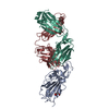
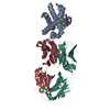
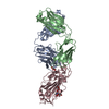
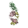
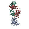
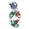
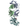
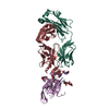
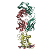
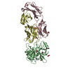
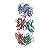
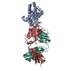
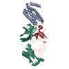
| Title | Structure of Hepatitis C Virus Envelope Glycoprotein E2 core from genotype 6a bound to broadly neutralizing antibody HC11 | |||||||||
 Components Components |
| |||||||||
 Keywords Keywords | IMMUNE SYSTEM / HCV / broadly neutralizing antibodies / bNAbs / E2 core / IGHV1-69 | |||||||||
| Function / homology |  Function and homology information Function and homology informationhost cell mitochondrial membrane / host cell lipid droplet / symbiont-mediated transformation of host cell / symbiont-mediated suppression of host TRAF-mediated signal transduction / symbiont-mediated perturbation of host cell cycle G1/S transition checkpoint / symbiont-mediated suppression of host JAK-STAT cascade via inhibition of STAT1 activity / symbiont-mediated suppression of host cytoplasmic pattern recognition receptor signaling pathway via inhibition of MAVS activity / ribonucleoside triphosphate phosphatase activity / viral nucleocapsid / channel activity ...host cell mitochondrial membrane / host cell lipid droplet / symbiont-mediated transformation of host cell / symbiont-mediated suppression of host TRAF-mediated signal transduction / symbiont-mediated perturbation of host cell cycle G1/S transition checkpoint / symbiont-mediated suppression of host JAK-STAT cascade via inhibition of STAT1 activity / symbiont-mediated suppression of host cytoplasmic pattern recognition receptor signaling pathway via inhibition of MAVS activity / ribonucleoside triphosphate phosphatase activity / viral nucleocapsid / channel activity / monoatomic ion transmembrane transport / clathrin-dependent endocytosis of virus by host cell / RNA helicase activity / host cell perinuclear region of cytoplasm / host cell endoplasmic reticulum membrane / symbiont-mediated suppression of host type I interferon-mediated signaling pathway / ribonucleoprotein complex / serine-type endopeptidase activity / symbiont-mediated activation of host autophagy / cysteine-type endopeptidase activity / viral RNA genome replication / RNA-directed RNA polymerase activity / fusion of virus membrane with host endosome membrane / viral envelope / virion attachment to host cell / host cell nucleus / host cell plasma membrane / virion membrane / structural molecule activity / proteolysis / RNA binding / zinc ion binding / ATP binding Similarity search - Function | |||||||||
| Biological species |  Homo sapiens (human) Homo sapiens (human) Recombinant Hepatitis C virus HK6a/JFH-1 Recombinant Hepatitis C virus HK6a/JFH-1 | |||||||||
| Method |  X-RAY DIFFRACTION / SYNCHROTRON / MOLECULAR REPLACEMENT / Resolution: 2.349 Å X-RAY DIFFRACTION / SYNCHROTRON / MOLECULAR REPLACEMENT / Resolution: 2.349 Å | |||||||||
 Authors Authors | Tzarum, N. / Wilson, I.A. / Law, M. | |||||||||
| Funding support |  United States, 2items United States, 2items
| |||||||||
 Citation Citation | Journal: Sci Adv / Year: 2020 Title: An alternate conformation of HCV E2 neutralizing face as an additional vaccine target. Authors: Tzarum, N. / Giang, E. / Kadam, R.U. / Chen, F. / Nagy, K. / Augestad, E.H. / Velazquez-Moctezuma, R. / Keck, Z.Y. / Hua, Y. / Stanfield, R.L. / Dreux, M. / Prentoe, J. / Foung, S.K.H. / ...Authors: Tzarum, N. / Giang, E. / Kadam, R.U. / Chen, F. / Nagy, K. / Augestad, E.H. / Velazquez-Moctezuma, R. / Keck, Z.Y. / Hua, Y. / Stanfield, R.L. / Dreux, M. / Prentoe, J. / Foung, S.K.H. / Bukh, J. / Wilson, I.A. / Law, M. | |||||||||
| History |
|
- Structure visualization
| Structure viewer | Molecule: MolmilJmol/JSmol |
|---|
- Downloads & links
-Download
| PDBx/mmCIF format | 6wo4.cif.gz | 135.4 KB | Display | PDBx/mmCIF format |
|---|---|---|---|---|
| PDB format | pdb6wo4.ent.gz | 102.1 KB | Display | PDB format |
| PDBx/mmJSON format | 6wo4.json.gz | Tree view | PDBx/mmJSON format | |
| Others |  Other downloads Other downloads |
-Validation report
| Arichive directory | https://data.pdbj.org/pub/pdb/validation_reports/wo/6wo4ftp://data.pdbj.org/pub/pdb/validation_reports/wo/6wo4 | HTTPS FTP |
|---|
-Related structure data
| Related structure data | 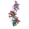 6wo3SC 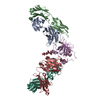 6wo5C 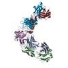 6woqC 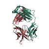 6worC  6wosC S: Starting model for refinement C: citing same article ( |
|---|---|
| Similar structure data |


-Links
 PDBj
PDBj


- Assembly
| Deposited unit | 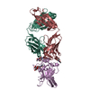
| ||||||||
|---|---|---|---|---|---|---|---|---|---|
| 1 |
| ||||||||
| Unit cell |
|
-Components
-Antibody , 2 types, 2 molecules HL
| #1: Antibody | Mass: 24164.342 Da / Num. of mol.: 1 Source method: isolated from a genetically manipulated source Source: (gene. exp.) Homo sapiens (human) / Production host: Homo sapiens (human) |
|---|---|
| #3: Antibody | Mass: 25430.373 Da / Num. of mol.: 1 Source method: isolated from a genetically manipulated source Source: (gene. exp.) Homo sapiens (human) / Production host: Homo sapiens (human) |
-Protein / Non-polymers , 2 types, 97 molecules E
| #2: Protein | Mass: 20707.395 Da / Num. of mol.: 1 Source method: isolated from a genetically manipulated source Source: (gene. exp.) Recombinant Hepatitis C virus HK6a/JFH-1Production host: Homo sapiens (human) / References: UniProt: B9V0E2*PLUS |
|---|---|
| #6: Water | ChemComp-HOH / Mass: 18.015 Da / Num. of mol.: 96 / Source method: isolated from a natural source / Formula: H2O |
-Sugars , 2 types, 4 molecules 
| #4: Polysaccharide | Source method: isolated from a genetically manipulated source #5: Sugar |  Type: D-saccharide, beta linking / Mass: 221.208 Da / Num. of mol.: 2 Type: D-saccharide, beta linking / Mass: 221.208 Da / Num. of mol.: 2Source method: isolated from a genetically manipulated source Formula: C8H15NO6 |
|---|
-Details
| Has ligand of interest | N |
|---|---|
| Has protein modification | Y |
-Experimental details
-Experiment
| Experiment | Method: X-RAY DIFFRACTION / Number of used crystals: 1 |
|---|
- Sample preparation
| Crystal | Density Matthews: 3.2 Å3/Da / Density % sol: 61.57 % |
|---|---|
| Crystal grow | Temperature: 293 K / Method: vapor diffusion, sitting drop / pH: 4.6 Details: 0.16 M ammonium sulfate, 20% (w/v) PEG 4000, 20% (v/v) glycerol, 0.08 M sodium acetate, pH 4.6 |
-Data collection
| Diffraction | Mean temperature: 100 K / Serial crystal experiment: N |
|---|---|
| Diffraction source | Source: SYNCHROTRON / Site: APS / Beamline: 23-ID-D / Wavelength: 1.0332 Å |
| Detector | Type: DECTRIS PILATUS3 S 6M / Detector: PIXEL / Date: Apr 7, 2018 |
| Radiation | Protocol: SINGLE WAVELENGTH / Monochromatic (M) / Laue (L): M / Scattering type: x-ray |
| Radiation wavelength | Wavelength: 1.0332 Å / Relative weight: 1 |
| Reflection | Resolution: 2.349→30 Å / Num. obs: 36627 / % possible obs: 94.5 % / Redundancy: 9.1 % / CC1/2: 0.92 / Net I/σ(I): 25.7 |
| Reflection shell | Resolution: 2.349→2.39 Å / Num. unique obs: 1693 / CC1/2: 0.57 |
- Processing
| Software |
| ||||||||||||||||||||||||||||||||||||||||||||||||||||||||||||||||||||||||||||||||||||
|---|---|---|---|---|---|---|---|---|---|---|---|---|---|---|---|---|---|---|---|---|---|---|---|---|---|---|---|---|---|---|---|---|---|---|---|---|---|---|---|---|---|---|---|---|---|---|---|---|---|---|---|---|---|---|---|---|---|---|---|---|---|---|---|---|---|---|---|---|---|---|---|---|---|---|---|---|---|---|---|---|---|---|---|---|---|
| Refinement | Method to determine structure: MOLECULAR REPLACEMENT Starting model: 6WO3 Resolution: 2.349→29.368 Å / SU ML: 0.35 / Cross valid method: THROUGHOUT / σ(F): 1.35 / Phase error: 25.79 / Stereochemistry target values: ML
| ||||||||||||||||||||||||||||||||||||||||||||||||||||||||||||||||||||||||||||||||||||
| Solvent computation | Shrinkage radii: 0.9 Å / VDW probe radii: 1.11 Å / Solvent model: FLAT BULK SOLVENT MODEL | ||||||||||||||||||||||||||||||||||||||||||||||||||||||||||||||||||||||||||||||||||||
| Displacement parameters | Biso max: 135.37 Å2 / Biso mean: 58.9708 Å2 / Biso min: 26.52 Å2 | ||||||||||||||||||||||||||||||||||||||||||||||||||||||||||||||||||||||||||||||||||||
| Refinement step | Cycle: final / Resolution: 2.349→29.368 Å
| ||||||||||||||||||||||||||||||||||||||||||||||||||||||||||||||||||||||||||||||||||||
| Refine LS restraints |
| ||||||||||||||||||||||||||||||||||||||||||||||||||||||||||||||||||||||||||||||||||||
| LS refinement shell | Refine-ID: X-RAY DIFFRACTION / Rfactor Rfree error: 0
|
