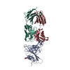
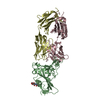
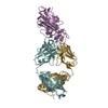
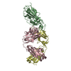
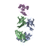
Hepatitis B virus e-antigen (HBeAg) is comprised of chains E and F / antibody Fab e6 fragment is comprised of two chains, one light (chains B or D) and one heavy (chains A or C). / one HBeAg-Fab complex involves one HBeAg and two Fabs, or the chains A-F of the crystal ASU.
-
Components
#1: Antibody
Fabe6HeavyChain
Mass: 23765.408 Da / Num. of mol.: 2 / Fragment: Fab e6 Heavy Chain / Source method: isolated from a natural source / Source: (natural) Mus musculus (house mouse)
#2: Antibody
Fabe6LightChain
Mass: 24329.928 Da / Num. of mol.: 2 / Fragment: Fab e6 Light Chain / Source method: isolated from a natural source / Source: (natural) Mus musculus (house mouse)
#3: Protein
e-antigen
Mass: 17879.451 Da / Num. of mol.: 2 / Fragment: HBV e-antigen (Cp(-10)149) / Mutation: C48A, C107A, G123A Source method: isolated from a genetically manipulated source Source: (gene. exp.) Hepatitis B virus / Gene: preC/C / References: UniProt: Q9QMH8
Has protein modification
Y
-
Experimental details
-
Experiment
Experiment
Method: X-RAY DIFFRACTION
-
Sample preparation
Crystal
Density Matthews: 2.86 Å3/Da / Density % sol: 57.05 %
Method to determine structure: MOLECULAR REPLACEMENT / Resolution: 3.34→46.2 Å / Cor.coef. Fo:Fc: 0.8793 / Cor.coef. Fo:Fc free: 0.8901 / Cross valid method: THROUGHOUT / σ(F): 0 Details: THE ROBS, RWORK AND RFREE VALUES ARE VERY CLOSE DUE TO THE USE OF TARGET RESTRAINTS DURING REFINEMENT FOR THE FAB MOLECULES TO A PREVIOUSLY-DETERMINED HIGHER RESOLUTION STRUCTURE (2.3 ...Details: THE ROBS, RWORK AND RFREE VALUES ARE VERY CLOSE DUE TO THE USE OF TARGET RESTRAINTS DURING REFINEMENT FOR THE FAB MOLECULES TO A PREVIOUSLY-DETERMINED HIGHER RESOLUTION STRUCTURE (2.3 ANGSTROM; PDB ID 3V6F) (SMART ET AL., 2012)
Rfactor
Num. reflection
% reflection
Selection details
Rfree
0.2333
1004
5.11 %
RANDOM
Rwork
0.234
-
-
-
obs
0.234
19651
92.62 %
-
Displacement parameters
Biso mean: 110.97 Å2
Baniso -1
Baniso -2
Baniso -3
1-
1.5488 Å2
-8.6258 Å2
-8.9658 Å2
2-
-
-6.3674 Å2
16.7917 Å2
3-
-
-
4.8185 Å2
Refine analyze
Luzzati coordinate error obs: 0.937 Å
Refinement step
Cycle: LAST / Resolution: 3.34→46.2 Å
Protein
Nucleic acid
Ligand
Solvent
Total
Num. atoms
9082
0
0
0
9082
Refine LS restraints
Refine-ID
Type
Dev ideal
Number
Restraint function
Weight
X-RAY DIFFRACTION
t_bond_d
0.008
9302
HARMONIC
2
X-RAY DIFFRACTION
t_angle_deg
1.19
12699
HARMONIC
2
X-RAY DIFFRACTION
t_dihedral_angle_d
3005
SINUSOIDAL
2
X-RAY DIFFRACTION
t_incorr_chiral_ct
X-RAY DIFFRACTION
t_pseud_angle
X-RAY DIFFRACTION
t_trig_c_planes
174
HARMONIC
2
X-RAY DIFFRACTION
t_gen_planes
1353
HARMONIC
5
X-RAY DIFFRACTION
t_it
9302
HARMONIC
20
X-RAY DIFFRACTION
t_nbd
X-RAY DIFFRACTION
t_omega_torsion
2.96
X-RAY DIFFRACTION
t_other_torsion
21.46
X-RAY DIFFRACTION
t_improper_torsion
X-RAY DIFFRACTION
t_chiral_improper_torsion
1242
SEMIHARMONIC
5
X-RAY DIFFRACTION
t_sum_occupancies
X-RAY DIFFRACTION
t_utility_distance
X-RAY DIFFRACTION
t_utility_angle
X-RAY DIFFRACTION
t_utility_torsion
X-RAY DIFFRACTION
t_ideal_dist_contact
10209
SEMIHARMONIC
4
LS refinement shell
Resolution: 3.34→3.52 Å / Total num. of bins used: 10
Rfactor
Num. reflection
% reflection
Rfree
0.2781
134
4.84 %
Rwork
0.246
2635
-
all
0.2475
2769
-
obs
-
-
92.62 %
Refinement TLS params.
Method: refined / Refine-ID: X-RAY DIFFRACTION
ID
L11 (°2)
L12 (°2)
L13 (°2)
L22 (°2)
L23 (°2)
L33 (°2)
S11 (Å °)
S12 (Å °)
S13 (Å °)
S21 (Å °)
S22 (Å °)
S23 (Å °)
S31 (Å °)
S32 (Å °)
S33 (Å °)
T11 (Å2)
T12 (Å2)
T13 (Å2)
T22 (Å2)
T23 (Å2)
T33 (Å2)
Origin x (Å)
Origin y (Å)
Origin z (Å)
1
8.1163
0.2308
1.6747
3.6032
0.5947
5.1577
0.1487
-0.2225
-0.3728
0.2763
-0.4463
0.3245
0.1821
-0.1336
0.2976
-0.0565
-0.0196
-0.0746
-0.4174
0.0079
0.2337
-5.8528
-8.0549
65.6961
2
5.51
-1.2686
0.8239
4.9691
-3.0023
1.9324
0.2379
-0.1962
0.1505
-0.312
-0.0363
0.1347
-0.6114
-0.0333
-0.2015
0.3081
0.1431
0.0554
-0.244
-0.0194
-0.1914
15.6472
17.0931
60.6451
3
8.1033
-1.9239
-1.7567
4.2318
1.3885
7.7908
0.2308
0.0284
0.1363
-0.1734
-0.4952
0.1667
-0.0535
-0.0473
0.2644
-0.0884
0.0125
-0.0575
-0.3638
0.0683
0.0152
-6.1585
-36.301
8.6852
4
7.4969
-0.8769
1.454
5.4521
-2.3813
2.6122
0.1449
0.104
-0.2805
0.0939
-0.0978
-0.0519
0.437
-0.4641
-0.0471
0.2753
0.1045
-0.0684
-0.2609
0.0811
-0.2131
15.4538
-61.3629
13.7987
5
5.273
1.8776
-2.3656
3.4947
0.2504
5.6164
-0.1877
0.6372
-0.2001
-0.1502
-0.2923
0.1856
0.2812
-0.1098
0.48
-0.2102
0.2259
-0.1301
-0.0697
-0.124
0.0782
-3.5084
-10.3966
44.5177
6
6.4548
0.1899
2.5055
5.2161
0.9064
8.9339
0.2509
-0.0798
-0.1085
-0.0338
-0.3233
-0.1464
-0.2838
0.3569
0.0724
-0.0135
-0.045
0.0961
-0.2736
0.1858
-0.1205
27.3521
12.3976
50.9139
7
3.8172
0.9301
2.1894
3.3999
0.0012
8.9825
-0.007
-0.7482
0.3027
0.1362
-0.1572
0.2442
0.0803
0.3002
0.1642
-0.2776
-0.1357
0.0594
-0.0981
-0.0461
0.0433
-3.5894
-33.6597
29.8124
8
9.5569
-1.1865
-2.9122
3.3454
-0.1102
6.8917
0.1254
-0.0251
-0.037
-0.0534
-0.1389
-0.2237
0.0922
0.501
0.0135
-0.017
0.2462
-0.0063
-0.1553
0.192
-0.1196
27.1767
-56.6172
23.3532
9
5.5111
-4.1621
-2.3648
2.9597
0.8174
2.4854
0.1889
-0.0998
0.2291
0.0382
0.0059
-0.0014
-0.1869
-0.4332
-0.1948
-0.41
0.1225
0.0318
-0.0926
-0.1609
0.2674
-35.6645
-23.019
20.0986
10
4.4414
4.4144
1.3269
4.0834
1.6038
1.1859
0.0872
0.0963
-0.1498
0.0496
0.0168
-0.1529
0.1282
-0.5412
-0.104
-0.4233
0.0313
0.0096
-0.0582
-0.1658
0.2732
-35.5085
-21.1849
54.3545
Refinement TLS group
ID
Refine-ID
Refine TLS-ID
Selection details
Auth asym-ID
Auth seq-ID
1
X-RAY DIFFRACTION
1
{A|1 - 123}
A
1 - 123
2
X-RAY DIFFRACTION
2
{A|124 - 224}
A
124 - 224
3
X-RAY DIFFRACTION
3
{C|1 - 123}
C
1 - 123
4
X-RAY DIFFRACTION
4
{C|124 - 224}
C
124 - 224
5
X-RAY DIFFRACTION
5
{B|1 - 115}
B
1 - 115
6
X-RAY DIFFRACTION
6
{B|116 - 219}
B
116 - 219
7
X-RAY DIFFRACTION
7
{D|1 - 115}
D
1 - 115
8
X-RAY DIFFRACTION
8
{D|116 - 219}
D
116 - 219
9
X-RAY DIFFRACTION
9
{E|4 - 151}
E
4 - 151
10
X-RAY DIFFRACTION
10
{F|4 - 151}
F
4 - 151
+
About Yorodumi
-
News
-
Feb 9, 2022. New format data for meta-information of EMDB entries
New format data for meta-information of EMDB entries
Version 3 of the EMDB header file is now the official format.
The previous official version 1.9 will be removed from the archive.
In the structure databanks used in Yorodumi, some data are registered as the other names, "COVID-19 virus" and "2019-nCoV". Here are the details of the virus and the list of structure data.
Jan 31, 2019. EMDB accession codes are about to change! (news from PDBe EMDB page)
EMDB accession codes are about to change! (news from PDBe EMDB page)
The allocation of 4 digits for EMDB accession codes will soon come to an end. Whilst these codes will remain in use, new EMDB accession codes will include an additional digit and will expand incrementally as the available range of codes is exhausted. The current 4-digit format prefixed with “EMD-” (i.e. EMD-XXXX) will advance to a 5-digit format (i.e. EMD-XXXXX), and so on. It is currently estimated that the 4-digit codes will be depleted around Spring 2019, at which point the 5-digit format will come into force.
The EM Navigator/Yorodumi systems omit the EMD- prefix.
Related info.:Q: What is EMD? / ID/Accession-code notation in Yorodumi/EM Navigator
Yorodumi is a browser for structure data from EMDB, PDB, SASBDB, etc.
This page is also the successor to EM Navigator detail page, and also detail information page/front-end page for Omokage search.
The word "yorodu" (or yorozu) is an old Japanese word meaning "ten thousand". "mi" (miru) is to see.
Related info.:EMDB / PDB / SASBDB / Comparison of 3 databanks / Yorodumi Search / Aug 31, 2016. New EM Navigator & Yorodumi / Yorodumi Papers / Jmol/JSmol / Function and homology information / Changes in new EM Navigator and Yorodumi
 Movie
Movie Controller


 Open data
 Basic information
 Components
 Keywords
 Function and homology information
 Hepatitis B virus
 X-RAY DIFFRACTION /
  Authors
 Citation
 Structure visualization
 Downloads & links
 Other downloads


 PDBj
PDBj


 Assembly
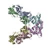


 Sample preparation
 / Beamline: I24
 Processing