 Movie
Movie Controller

[English] 日本語
 Yorodumi
- PDB-6xy2: Crystal structure of CTLA-4 complexed with the Fab of HL32 antibody -
+ Open data

- Basic information
| Entry | Database: PDB / ID: 6xy2 | ||||||
|---|---|---|---|---|---|---|---|
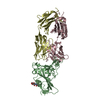
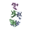
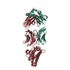
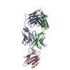
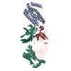
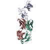
| Title | Crystal structure of CTLA-4 complexed with the Fab of HL32 antibody | ||||||
 Components Components |
| ||||||
 Keywords Keywords | IMMUNE SYSTEM / CTLA-4 / Fab / antibody / immunnotherapy | ||||||
| Function / homology |  Function and homology information Function and homology informationreceptor decoy activity / protein complex involved in cell adhesion / RUNX1 and FOXP3 control the development of regulatory T lymphocytes (Tregs) / clathrin-coated endocytic vesicle / Co-stimulation by CD28 / negative regulation of regulatory T cell differentiation / Co-inhibition by CTLA4 / negative regulation of T cell activation / negative regulation of B cell proliferation / negative regulation of T cell receptor signaling pathway ...receptor decoy activity / protein complex involved in cell adhesion / RUNX1 and FOXP3 control the development of regulatory T lymphocytes (Tregs) / clathrin-coated endocytic vesicle / Co-stimulation by CD28 / negative regulation of regulatory T cell differentiation / Co-inhibition by CTLA4 / negative regulation of T cell activation / negative regulation of B cell proliferation / negative regulation of T cell receptor signaling pathway / negative regulation of T cell proliferation / B cell receptor signaling pathway / T cell receptor signaling pathway / adaptive immune response / immune response / positive regulation of apoptotic process / external side of plasma membrane / DNA damage response / perinuclear region of cytoplasm / Golgi apparatus / plasma membrane Similarity search - Function | ||||||
| Biological species |  Homo sapiens (human) Homo sapiens (human) | ||||||
| Method |  X-RAY DIFFRACTION / SYNCHROTRON / MOLECULAR REPLACEMENT / Resolution: 3.05 Å X-RAY DIFFRACTION / SYNCHROTRON / MOLECULAR REPLACEMENT / Resolution: 3.05 Å | ||||||
 Authors Authors | Gao, H. / Zhou, A. | ||||||
| Funding support |  China, 1items China, 1items
| ||||||
 Citation Citation | Journal: Cell Discov / Year: 2020 Title: Structure of CTLA-4 complexed with a pH-sensitive cancer immunotherapeutic antibody. Authors: Gao, H. / Cai, H. / Liu, J. / Wang, X. / Zheng, P. / Devenport, M. / Xu, T. / Dou, F. / Liu, Y. / Zhou, A. | ||||||
| History |
|
- Structure visualization
| Structure viewer | Molecule: MolmilJmol/JSmol |
|---|
- Downloads & links
-Download
| PDBx/mmCIF format | 6xy2.cif.gz | 374.3 KB | Display | PDBx/mmCIF format |
|---|---|---|---|---|
| PDB format | pdb6xy2.ent.gz | 261.1 KB | Display | PDB format |
| PDBx/mmJSON format | 6xy2.json.gz | Tree view | PDBx/mmJSON format | |
| Others |  Other downloads Other downloads |
-Validation report
| Arichive directory | https://data.pdbj.org/pub/pdb/validation_reports/xy/6xy2ftp://data.pdbj.org/pub/pdb/validation_reports/xy/6xy2 | HTTPS FTP |
|---|
-Related structure data
| Related structure data |  3oskS S: Starting model for refinement |
|---|---|
| Similar structure data |


-Links
 PDBj
PDBj


- Assembly
| Deposited unit | 
| ||||||||||
|---|---|---|---|---|---|---|---|---|---|---|---|
| 1 |
| ||||||||||
| Unit cell |
|
-Components
| #1: Protein | Mass: 13194.995 Da / Num. of mol.: 1 Source method: isolated from a genetically manipulated source Source: (gene. exp.) Homo sapiens (human) / Gene: CTLA4, CD152 / Cell line (production host): HEK293 / Production host: Homo sapiens (human) / References: UniProt: P16410 |
|---|---|
| #2: Antibody | Mass: 26582.570 Da / Num. of mol.: 1 Source method: isolated from a genetically manipulated source Source: (gene. exp.) Homo sapiens (human) / Cell line (production host): HEK293 / Production host: Homo sapiens (human) |
| #3: Antibody | Mass: 23505.047 Da / Num. of mol.: 1 Source method: isolated from a genetically manipulated source Source: (gene. exp.) Homo sapiens (human) / Cell line (production host): HEK293 / Production host: Homo sapiens (human) |
| #4: Sugar | ChemComp-NAG /   Type: D-saccharide, beta linking / Mass: 221.208 Da / Num. of mol.: 1 Type: D-saccharide, beta linking / Mass: 221.208 Da / Num. of mol.: 1Source method: isolated from a genetically manipulated source Formula: C8H15NO6 |
| Has ligand of interest | N |
| Has protein modification | Y |
-Experimental details
-Experiment
| Experiment | Method: X-RAY DIFFRACTION / Number of used crystals: 1 |
|---|
- Sample preparation
| Crystal | Density Matthews: 5.5 Å3/Da / Density % sol: 77.62 % |
|---|---|
| Crystal grow | Temperature: 293 K / Method: batch mode / pH: 6.5 / Details: 1M Li2SO4, 1.5M NH4SO4, 0.1M Sodium citrate |
-Data collection
| Diffraction | Mean temperature: 100 K / Serial crystal experiment: N |
|---|---|
| Diffraction source | Source: SYNCHROTRON / Site: SSRF / Beamline: BL19U1 / Wavelength: 0.97892 Å |
| Detector | Type: DECTRIS PILATUS3 S 6M / Detector: PIXEL / Date: Jan 13, 2020 |
| Radiation | Protocol: SINGLE WAVELENGTH / Monochromatic (M) / Laue (L): M / Scattering type: x-ray |
| Radiation wavelength | Wavelength: 0.97892 Å / Relative weight: 1 |
| Reflection | Resolution: 2.95→85 Å / Num. obs: 29874 / % possible obs: 99.9 % / Redundancy: 7.9 % / Biso Wilson estimate: 100.72 Å2 / CC1/2: 0.992 / Rmerge(I) obs: 0.133 / Rpim(I) all: 0.049 / Rrim(I) all: 0.142 / Net I/σ(I): 7.4 |
| Reflection shell | Resolution: 2.95→3.11 Å / Rmerge(I) obs: 1.865 / Mean I/σ(I) obs: 0.7 / Num. unique obs: 4226 / CC1/2: 0.372 / Rpim(I) all: 0.772 |
- Processing
| Software |
| |||||||||||||||||||||||||||||||||||||||||||||||||||||||||||||||||||||||||||||
|---|---|---|---|---|---|---|---|---|---|---|---|---|---|---|---|---|---|---|---|---|---|---|---|---|---|---|---|---|---|---|---|---|---|---|---|---|---|---|---|---|---|---|---|---|---|---|---|---|---|---|---|---|---|---|---|---|---|---|---|---|---|---|---|---|---|---|---|---|---|---|---|---|---|---|---|---|---|---|
| Refinement | Method to determine structure: MOLECULAR REPLACEMENT Starting model: 3OSK Resolution: 3.05→75.33 Å / SU ML: 0.5355 / Cross valid method: FREE R-VALUE / σ(F): 1.34 / Phase error: 35.7336 Stereochemistry target values: GeoStd + Monomer Library + CDL v1.2
| |||||||||||||||||||||||||||||||||||||||||||||||||||||||||||||||||||||||||||||
| Solvent computation | Shrinkage radii: 0.9 Å / VDW probe radii: 1.11 Å / Solvent model: FLAT BULK SOLVENT MODEL | |||||||||||||||||||||||||||||||||||||||||||||||||||||||||||||||||||||||||||||
| Displacement parameters | Biso mean: 70 Å2 | |||||||||||||||||||||||||||||||||||||||||||||||||||||||||||||||||||||||||||||
| Refinement step | Cycle: LAST / Resolution: 3.05→75.33 Å
| |||||||||||||||||||||||||||||||||||||||||||||||||||||||||||||||||||||||||||||
| Refine LS restraints |
| |||||||||||||||||||||||||||||||||||||||||||||||||||||||||||||||||||||||||||||
| LS refinement shell |
| |||||||||||||||||||||||||||||||||||||||||||||||||||||||||||||||||||||||||||||
| Refinement TLS params. | Method: refined / Origin x: 28.2864927151 Å / Origin y: 32.5358881788 Å / Origin z: -10.0469404765 Å
| |||||||||||||||||||||||||||||||||||||||||||||||||||||||||||||||||||||||||||||
| Refinement TLS group | Selection details: all |
