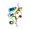
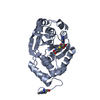
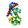
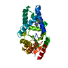
- PDB-6wh2: Structure of the C-terminal BRCT domain of human XRCC1 -
+
Open data
ID or keywords:
Loading...
-
Basic information
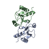
Entry
Database: PDB / ID: 6wh2
Title
Structure of the C-terminal BRCT domain of human XRCC1
Components
X-ray repair cross complementing protein 1 variant
Keywords
DNA BINDING PROTEIN / DNA ligase complex / DNA repair
Function / homology
Function and homology information
oxidized DNA binding / telomeric DNA-containing double minutes formation / ERCC4-ERCC1 complex / negative regulation of protection from non-homologous end joining at telomere / ADP-D-ribose modification-dependent protein binding / negative regulation of protein ADP-ribosylation / poly-ADP-D-ribose binding / regulation of base-excision repair / single strand break repair / HDR through MMEJ (alt-NHEJ) ...oxidized DNA binding / telomeric DNA-containing double minutes formation / ERCC4-ERCC1 complex / negative regulation of protection from non-homologous end joining at telomere / ADP-D-ribose modification-dependent protein binding / negative regulation of protein ADP-ribosylation / poly-ADP-D-ribose binding / regulation of base-excision repair / single strand break repair / HDR through MMEJ (alt-NHEJ) / 3' overhang single-stranded DNA endonuclease activity / response to hydroperoxide / Resolution of AP sites via the single-nucleotide replacement pathway / APEX1-Independent Resolution of AP Sites via the Single Nucleotide Replacement Pathway / site of DNA damage / Gap-filling DNA repair synthesis and ligation in GG-NER / hippocampus development / base-excision repair / double-strand break repair via nonhomologous end joining / Gap-filling DNA repair synthesis and ligation in TC-NER / double-strand break repair / chromosome / damaged DNA binding / chromosome, telomeric region / chromatin / nucleolus / enzyme binding / nucleoplasm / nucleus Similarity search - Function
DNA-repair protein Xrcc1, N-terminal / XRCC1, first (central) BRCT domain / XRCC1 N terminal domain / BRCT domain / BRCA1 C Terminus (BRCT) domain / breast cancer carboxy-terminal domain / BRCT domain profile. / BRCT domain / BRCT domain superfamily / Galactose-binding-like domain superfamily Similarity search - Domain/homology
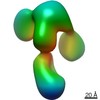
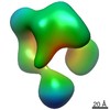
Journal: Nucleic Acids Res / Year: 2021 Title: An atypical BRCT-BRCT interaction with the XRCC1 scaffold protein compacts human DNA Ligase IIIα within a flexible DNA repair complex. Authors: Michal Hammel / Ishtiaque Rashid / Aleksandr Sverzhinsky / Yasin Pourfarjam / Miaw-Sheue Tsai / Tom Ellenberger / John M Pascal / In-Kwon Kim / John A Tainer / Alan E Tomkinson / Abstract: The XRCC1-DNA ligase IIIα complex (XL) is critical for DNA single-strand break repair, a key target for PARP inhibitors in cancer cells deficient in homologous recombination. Here, we combined ...The XRCC1-DNA ligase IIIα complex (XL) is critical for DNA single-strand break repair, a key target for PARP inhibitors in cancer cells deficient in homologous recombination. Here, we combined biophysical approaches to gain insights into the shape and conformational flexibility of the XL as well as XRCC1 and DNA ligase IIIα (LigIIIα) alone. Structurally-guided mutational analyses based on the crystal structure of the human BRCT-BRCT heterodimer identified the network of salt bridges that together with the N-terminal extension of the XRCC1 C-terminal BRCT domain constitute the XL molecular interface. Coupling size exclusion chromatography with small angle X-ray scattering and multiangle light scattering (SEC-SAXS-MALS), we determined that the XL is more compact than either XRCC1 or LigIIIα, both of which form transient homodimers and are highly disordered. The reduced disorder and flexibility allowed us to build models of XL particles visualized by negative stain electron microscopy that predict close spatial organization between the LigIIIα catalytic core and both BRCT domains of XRCC1. Together our results identify an atypical BRCT-BRCT interaction as the stable nucleating core of the XL that links the flexible nick sensing and catalytic domains of LigIIIα to other protein partners of the flexible XRCC1 scaffold.
Mass: 11419.856 Da / Num. of mol.: 2 / Fragment: Second BRCT domain Source method: isolated from a genetically manipulated source Source: (gene. exp.) Homo sapiens (human) / Production host: Escherichia coli (E. coli) / References: UniProt: Q59HH7, UniProt: P18887*PLUS
In the structure databanks used in Yorodumi, some data are registered as the other names, "COVID-19 virus" and "2019-nCoV". Here are the details of the virus and the list of structure data.
Jan 31, 2019. EMDB accession codes are about to change! (news from PDBe EMDB page)
EMDB accession codes are about to change! (news from PDBe EMDB page)
The allocation of 4 digits for EMDB accession codes will soon come to an end. Whilst these codes will remain in use, new EMDB accession codes will include an additional digit and will expand incrementally as the available range of codes is exhausted. The current 4-digit format prefixed with “EMD-” (i.e. EMD-XXXX) will advance to a 5-digit format (i.e. EMD-XXXXX), and so on. It is currently estimated that the 4-digit codes will be depleted around Spring 2019, at which point the 5-digit format will come into force.
The EM Navigator/Yorodumi systems omit the EMD- prefix.
Related info.:Q: What is EMD? / ID/Accession-code notation in Yorodumi/EM Navigator
Yorodumi is a browser for structure data from EMDB, PDB, SASBDB, etc.
This page is also the successor to EM Navigator detail page, and also detail information page/front-end page for Omokage search.
The word "yorodu" (or yorozu) is an old Japanese word meaning "ten thousand". "mi" (miru) is to see.
Related info.:EMDB / PDB / SASBDB / Comparison of 3 databanks / Yorodumi Search / Aug 31, 2016. New EM Navigator & Yorodumi / Yorodumi Papers / Jmol/JSmol / Function and homology information / Changes in new EM Navigator and Yorodumi
 Movie
Movie Controller


 Open data
 Basic information
 Components
 Keywords
 Function and homology information
 Homo sapiens (human)
 X-RAY DIFFRACTION /
  Authors
 United States, 1items
  Citation
 Structure visualization
 Downloads & links
 Other downloads


 PDBj
PDBj


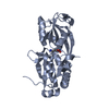
 Assembly
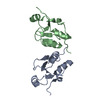

 Mass: 18.015 Da / Num. of mol.: 53 / Source method: isolated from a natural source / Formula: H2O
 Sample preparation
 Processing