Resolution: 2.6→2.69 Å / Redundancy: 5.1 % / Rmerge(I) obs: 0.676 / Mean I/σ(I) obs: 1.98 / Num. unique all: 2774 / % possible all: 96.7
-
Processing
Software
Name
Version
Classification
HKL-2000
datacollection
SnB
phasing
PHENIX
(phenix.refine: 1.6_289)
refinement
DENZO
datareduction
SCALEPACK
datascaling
Refinement
Method to determine structure: SAD / Resolution: 2.601→34.402 Å / SU ML: 0.25 / Cross valid method: THROUGHOUT / σ(F): 1.34 / Stereochemistry target values: ML
Rfactor
Num. reflection
% reflection
Selection details
Rfree
0.2783
360
4.6 %
RANDOM
Rwork
0.2372
-
-
-
obs
0.239
7832
98.35 %
-
Solvent computation
Shrinkage radii: 0.9 Å / VDW probe radii: 1.11 Å / Solvent model: FLAT BULK SOLVENT MODEL / Bsol: 64.356 Å2 / ksol: 0.348 e/Å3
Refinement step
Cycle: LAST / Resolution: 2.601→34.402 Å
Protein
Nucleic acid
Ligand
Solvent
Total
Num. atoms
1670
0
0
18
1688
Refine LS restraints
Refine-ID
Type
Dev ideal
Number
X-RAY DIFFRACTION
f_bond_d
0.009
1702
X-RAY DIFFRACTION
f_angle_d
1.566
2290
X-RAY DIFFRACTION
f_dihedral_angle_d
18.613
644
X-RAY DIFFRACTION
f_chiral_restr
0.097
263
X-RAY DIFFRACTION
f_plane_restr
0.006
283
LS refinement shell
Resolution (Å)
Rfactor Rfree
Num. reflection Rfree
Rfactor Rwork
Num. reflection Rwork
Refine-ID
% reflection obs (%)
2.6007-2.9768
0.4118
108
0.3491
2411
X-RAY DIFFRACTION
99
2.9768-3.7498
0.2865
117
0.2328
2478
X-RAY DIFFRACTION
99
3.7498-34.4048
0.2494
135
0.2129
2583
X-RAY DIFFRACTION
97
Refinement TLS params.
Refine-ID: X-RAY DIFFRACTION
ID
Method
L11 (°2)
L12 (°2)
L13 (°2)
L22 (°2)
L23 (°2)
L33 (°2)
S11 (Å °)
S12 (Å °)
S13 (Å °)
S21 (Å °)
S22 (Å °)
S23 (Å °)
S31 (Å °)
S32 (Å °)
S33 (Å °)
T11 (Å2)
T12 (Å2)
T13 (Å2)
T22 (Å2)
T23 (Å2)
T33 (Å2)
Origin x (Å)
Origin y (Å)
Origin z (Å)
1
refined
2.2411
1.6598
0.0948
-0.3716
1.1599
1.8668
0.0055
0.1209
0.0451
0.0006
-0.2214
-0.0411
-0.2671
-0.2313
0
0.438
0.0326
-0.0535
0.4507
-0.0397
0.4355
7.9845
20.995
61.1311
2
-0.0476
0.2818
-0.2595
0.7424
0.8945
1.6141
-0.1306
0.1772
-0.1806
-0.0169
0.1048
0.069
-0.3791
0.1825
-0
0.4845
-0.0678
-0.0625
0.5077
0.0069
0.4072
Refinement TLS group
Selection details: chain B
+
About Yorodumi
-
News
-
Feb 9, 2022. New format data for meta-information of EMDB entries
New format data for meta-information of EMDB entries
Version 3 of the EMDB header file is now the official format.
The previous official version 1.9 will be removed from the archive.
In the structure databanks used in Yorodumi, some data are registered as the other names, "COVID-19 virus" and "2019-nCoV". Here are the details of the virus and the list of structure data.
Jan 31, 2019. EMDB accession codes are about to change! (news from PDBe EMDB page)
EMDB accession codes are about to change! (news from PDBe EMDB page)
The allocation of 4 digits for EMDB accession codes will soon come to an end. Whilst these codes will remain in use, new EMDB accession codes will include an additional digit and will expand incrementally as the available range of codes is exhausted. The current 4-digit format prefixed with “EMD-” (i.e. EMD-XXXX) will advance to a 5-digit format (i.e. EMD-XXXXX), and so on. It is currently estimated that the 4-digit codes will be depleted around Spring 2019, at which point the 5-digit format will come into force.
The EM Navigator/Yorodumi systems omit the EMD- prefix.
Related info.:Q: What is EMD? / ID/Accession-code notation in Yorodumi/EM Navigator
Yorodumi is a browser for structure data from EMDB, PDB, SASBDB, etc.
This page is also the successor to EM Navigator detail page, and also detail information page/front-end page for Omokage search.
The word "yorodu" (or yorozu) is an old Japanese word meaning "ten thousand". "mi" (miru) is to see.
Related info.:EMDB / PDB / SASBDB / Comparison of 3 databanks / Yorodumi Search / Aug 31, 2016. New EM Navigator & Yorodumi / Yorodumi Papers / Jmol/JSmol / Function and homology information / Changes in new EM Navigator and Yorodumi
 Movie
Movie Controller

 Yorodumi
 Open data
 Basic information
 Components
 Keywords
 Function and homology information
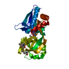
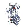
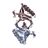
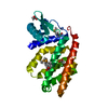
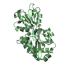
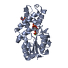
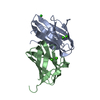
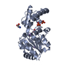
 Desulfitobacterium hafniense (bacteria)
 X-RAY DIFFRACTION /
  Authors
 Citation
 Structure visualization
 Downloads & links
 Other downloads
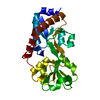

 PDBj
PDBj


 Assembly
 Mass: 18.015 Da / Num. of mol.: 18 / Source method: isolated from a natural source / Formula: H2O
 Sample preparation
 / Beamline: X4C / Wavelength: 0.979 Å
 Processing