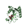
 Movie
Movie Controller


+ Open data

- Basic information
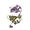
| Entry | Database: PDB / ID: 1i7a | ||||||
|---|---|---|---|---|---|---|---|
| Title | EVH1 DOMAIN FROM MURINE HOMER 2B/VESL 2 | ||||||
 Components Components |
| ||||||
 Keywords Keywords | SIGNALING PROTEIN / EVH1 domain / Homer / Vesl / X-ray crystal structure / brain | ||||||
| Function / homology |  Function and homology information Function and homology informationG protein-coupled glutamate receptor binding / Neurexins and neuroligins / synaptic receptor adaptor activity / regulation of glutamate secretion / regulation of store-operated calcium entry / stereocilium tip / G protein-coupled glutamate receptor signaling pathway / regulation of G protein-coupled receptor signaling pathway / negative regulation of calcineurin-NFAT signaling cascade / negative regulation of interleukin-2 production ...G protein-coupled glutamate receptor binding / Neurexins and neuroligins / synaptic receptor adaptor activity / regulation of glutamate secretion / regulation of store-operated calcium entry / stereocilium tip / G protein-coupled glutamate receptor signaling pathway / regulation of G protein-coupled receptor signaling pathway / negative regulation of calcineurin-NFAT signaling cascade / negative regulation of interleukin-2 production / glutamate receptor binding / regulation of synaptic transmission, glutamatergic / behavioral response to cocaine / calcium-mediated signaling / response to cocaine / sensory perception of sound / apical part of cell / actin binding / postsynaptic density / protein domain specific binding / neuronal cell body / dendrite / protein-containing complex binding / glutamatergic synapse / identical protein binding / plasma membrane / cytosol / cytoplasm Similarity search - Function | ||||||
| Biological species |  | ||||||
| Method |  X-RAY DIFFRACTION / MOLECULAR REPLACEMENT / Resolution: 2.24 Å X-RAY DIFFRACTION / MOLECULAR REPLACEMENT / Resolution: 2.24 Å | ||||||
 Authors Authors | Barzik, M. / Carl, U.D. / Schubert, W.-D. / Wehland, J. / Heinz, D.W. | ||||||
 Citation Citation | Journal: J.Mol.Biol. / Year: 2001 Title: The N-terminal domain of Homer/Vesl is a new class II EVH1 domain. Authors: Barzik, M. / Carl, U.D. / Schubert, W.D. / Frank, R. / Wehland, J. / Heinz, D.W. | ||||||
| History |
|
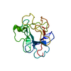
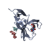
- Structure visualization
| Structure viewer | Molecule: MolmilJmol/JSmol |
|---|
- Downloads & links
-Download
| PDBx/mmCIF format | 1i7a.cif.gz | 97.2 KB | Display | PDBx/mmCIF format |
|---|---|---|---|---|
| PDB format | pdb1i7a.ent.gz | 75.7 KB | Display | PDB format |
| PDBx/mmJSON format | 1i7a.json.gz | Tree view | PDBx/mmJSON format | |
| Others |  Other downloads Other downloads |
-Validation report
| Arichive directory | https://data.pdbj.org/pub/pdb/validation_reports/i7/1i7aftp://data.pdbj.org/pub/pdb/validation_reports/i7/1i7a | HTTPS FTP |
|---|
-Related structure data
| Related structure data |  1ddwS S: Starting model for refinement |
|---|---|
| Similar structure data |


-Links
 PDBj
PDBj


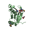
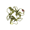
- Assembly
| Deposited unit | 
| ||||||||
|---|---|---|---|---|---|---|---|---|---|
| 1 | 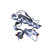
| ||||||||
| 2 | 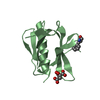
| ||||||||
| 3 | 
| ||||||||
| 4 | 
| ||||||||
| 5 | 
| ||||||||
| 6 | 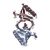
| ||||||||
| 7 | 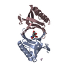
| ||||||||
| 8 | 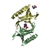
| ||||||||
| 9 | 
| ||||||||
| 10 | 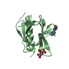
| ||||||||
| 11 | 
| ||||||||
| Unit cell |
|
-Components
| #1: Protein | Mass: 12576.193 Da / Num. of mol.: 4 / Fragment: EVH1 DOMAIN (N-TERMINAL) Source method: isolated from a genetically manipulated source Source: (gene. exp.)  #2: Protein/peptide | | Mass: 383.441 Da / Num. of mol.: 1 / Source method: isolated from a natural source / Source: (natural) #3: Chemical |   Mass: 189.100 Da / Num. of mol.: 2 / Source method: obtained synthetically / Formula: C6H5O7 Mass: 189.100 Da / Num. of mol.: 2 / Source method: obtained synthetically / Formula: C6H5O7#4: Chemical |   Type: L-peptide linking / Mass: 165.189 Da / Num. of mol.: 3 / Source method: obtained synthetically / Formula: C9H11NO2 Type: L-peptide linking / Mass: 165.189 Da / Num. of mol.: 3 / Source method: obtained synthetically / Formula: C9H11NO2#5: Water | ChemComp-HOH / |  Mass: 18.015 Da / Num. of mol.: 106 / Source method: isolated from a natural source / Formula: H2O Mass: 18.015 Da / Num. of mol.: 106 / Source method: isolated from a natural source / Formula: H2O |
|---|
-Experimental details
-Experiment
| Experiment | Method: X-RAY DIFFRACTION / Number of used crystals: 1 |
|---|
- Sample preparation
| Crystal | Density Matthews: 2.5 Å3/Da / Density % sol: 49 % | ||||||||||||||||||||
|---|---|---|---|---|---|---|---|---|---|---|---|---|---|---|---|---|---|---|---|---|---|
| Crystal grow | Temperature: 293 K / Method: vapor diffusion, hanging drop / pH: 7.5 Details: 1 M SODIUM CITRATE, O.1 M CHES, pH 9.8, pH 7.5, VAPOR DIFFUSION, HANGING DROP, temperature 293K | ||||||||||||||||||||
| Crystal | *PLUS Density % sol: 55.3 % | ||||||||||||||||||||
| Crystal grow | *PLUS Method: unknown | ||||||||||||||||||||
| Components of the solutions | *PLUS
|
-Data collection
| Diffraction | Mean temperature: 100 K |
|---|---|
| Diffraction source | Source: ROTATING ANODE / Type: RIGAKU RU200 / Wavelength: 1.5418 Å |
| Detector | Type: RIGAKU RAXIS IV / Detector: IMAGE PLATE / Date: Sep 15, 2000 / Details: mirrors |
| Radiation | Monochromator: mirrors / Protocol: SINGLE WAVELENGTH / Monochromatic (M) / Laue (L): M / Scattering type: x-ray |
| Radiation wavelength | Wavelength: 1.5418 Å / Relative weight: 1 |
| Reflection | Resolution: 2.2→40 Å / Num. all: 59066 / Num. obs: 22446 / % possible obs: 95.2 % / Observed criterion σ(F): 3 / Observed criterion σ(I): 3 / Redundancy: 2.6 % / Biso Wilson estimate: 45.8 Å2 / Rmerge(I) obs: 0.057 / Net I/σ(I): 15.4 |
| Reflection shell | Resolution: 2.2→2.28 Å / Redundancy: 2.1 % / Rmerge(I) obs: 0.28 / Mean I/σ(I) obs: 3 / Num. unique all: 2060 / % possible all: 88.2 |
| Reflection shell | *PLUS % possible obs: 88.2 % / Rmerge(I) obs: 0.28 |
- Processing
| Software |
| |||||||||||||||||||||||||
|---|---|---|---|---|---|---|---|---|---|---|---|---|---|---|---|---|---|---|---|---|---|---|---|---|---|---|
| Refinement | Method to determine structure: MOLECULAR REPLACEMENT Starting model: PDB ENTRY 1DDW (SAME MOLECULE IN DIFFERENT PACKING DERIVED FROM HOMER 1B FROM RAT Resolution: 2.24→15 Å / Isotropic thermal model: Isotropic / σ(F): 3 / σ(I): 1 / Stereochemistry target values: Engh & Huber / Details: The crystal had a particularly high mosaicity.
| |||||||||||||||||||||||||
| Displacement parameters |
| |||||||||||||||||||||||||
| Refinement step | Cycle: LAST / Resolution: 2.24→15 Å
| |||||||||||||||||||||||||
| Refine LS restraints |
| |||||||||||||||||||||||||
| LS refinement shell | Resolution: 2.2→2.24 Å
| |||||||||||||||||||||||||
| Software | *PLUS Name: CNS/REFMAC / Classification: refinement | |||||||||||||||||||||||||
| Refinement | *PLUS σ(F): 3 / % reflection Rfree: 4.9 % / Rfactor obs: 0.235 | |||||||||||||||||||||||||
| Solvent computation | *PLUS | |||||||||||||||||||||||||
| Displacement parameters | *PLUS | |||||||||||||||||||||||||
| Refine LS restraints | *PLUS
|
