 Movie
Movie Controller


+ Open data

- Basic information
| Entry | Database: PDB / ID: 6wb7 | ||||||
|---|---|---|---|---|---|---|---|
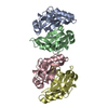
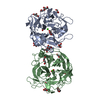
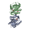
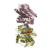
| Title | Acarbose Kinase AcbK as a Complex with Acarbose and AMP-PNP | ||||||
 Components Components | Acarbose 7(IV)-phosphotransferase | ||||||
 Keywords Keywords | TRANSFERASE / CARBOHYDRATE KINASE / ACARBOSE / NUCLEOTIDE BINDING / ATP BINDING / METAL ION BINDING / RIBOKINASE ACTIVITY / COMPLEX | ||||||
| Function / homology |  Function and homology information Function and homology information | ||||||
| Biological species |  Actinoplanes sp. (bacteria) Actinoplanes sp. (bacteria) | ||||||
| Method |  X-RAY DIFFRACTION / SYNCHROTRON / MOLECULAR REPLACEMENT / molecular replacement / Resolution: 2.44 Å X-RAY DIFFRACTION / SYNCHROTRON / MOLECULAR REPLACEMENT / molecular replacement / Resolution: 2.44 Å | ||||||
 Authors Authors | Jeffrey, P.D. / Balaich, J.N. / Estrella, M.A. / Donia, M.S. | ||||||
| Funding support | 1items
| ||||||
 Citation Citation | Journal: Nature / Year: 2021 Title: The human microbiome encodes resistance to the antidiabetic drug acarbose. Authors: Balaich, J. / Estrella, M. / Wu, G. / Jeffrey, P.D. / Biswas, A. / Zhao, L. / Korennykh, A. / Donia, M.S. | ||||||
| History |
|
- Structure visualization
| Structure viewer | Molecule: MolmilJmol/JSmol |
|---|
- Downloads & links
-Download
| PDBx/mmCIF format | 6wb7.cif.gz | 245.8 KB | Display | PDBx/mmCIF format |
|---|---|---|---|---|
| PDB format | pdb6wb7.ent.gz | 194.6 KB | Display | PDB format |
| PDBx/mmJSON format | 6wb7.json.gz | Tree view | PDBx/mmJSON format | |
| Others |  Other downloads Other downloads |
-Validation report
| Arichive directory | https://data.pdbj.org/pub/pdb/validation_reports/wb/6wb7ftp://data.pdbj.org/pub/pdb/validation_reports/wb/6wb7 | HTTPS FTP |
|---|
-Related structure data
| Related structure data |  6wb4SC  6wb5C S: Starting model for refinement C: citing same article ( |
|---|---|
| Similar structure data |


-Links
 PDBj
PDBj


- Assembly
| Deposited unit | 
| ||||||||
|---|---|---|---|---|---|---|---|---|---|
| 1 | 
| ||||||||
| 2 | 
| ||||||||
| Unit cell |
|
-Components
-Protein / Sugars , 2 types, 8 molecules ABCD
| #1: Protein | Mass: 33686.797 Da / Num. of mol.: 4 Source method: isolated from a genetically manipulated source Source: (gene. exp.) Actinoplanes sp. (strain ATCC 31044 / CBS 674.73 / SE50/110) (bacteria)Strain: ATCC 31044 / CBS 674.73 / SE50/110 / Gene: acbK, ACPL_3675 / Plasmid: pET28a / Production host: References: UniProt: Q8RMD4, acarbose 7IV-phosphotransferase #2: Polysaccharide | 4,6-dideoxy-4-{[(1S,4R,5S,6S)-4,5,6-trihydroxy-3-(hydroxymethyl)cyclohex-2-en-1-yl]amino}-alpha-D- ...4,6-dideoxy-4-{[(1S,4R,5S,6S)-4,5,6-trihydroxy-3-(hydroxymethyl)cyclohex-2-en-1-yl]amino}-alpha-D-glucopyranose-(1-4)-alpha-D-glucopyranose-(1-4)-alpha-D-glucopyranose / alpha-acarbose   Type: oligosaccharide, Oligosaccharide / Class: Inhibitor / Mass: 645.606 Da / Num. of mol.: 4 Type: oligosaccharide, Oligosaccharide / Class: Inhibitor / Mass: 645.606 Da / Num. of mol.: 4Source method: isolated from a genetically manipulated source Details: oligosaccharide / References: alpha-acarbose |
|---|
-Non-polymers , 4 types, 393 molecules 


| #3: Chemical | ChemComp-ANP /  Mass: 506.196 Da / Num. of mol.: 4 / Source method: obtained synthetically / Formula: C10H17N6O12P3 / Comment: AMP-PNP, energy-carrying molecule analogue*YM Mass: 506.196 Da / Num. of mol.: 4 / Source method: obtained synthetically / Formula: C10H17N6O12P3 / Comment: AMP-PNP, energy-carrying molecule analogue*YM#4: Chemical | ChemComp-MN /  Mass: 54.938 Da / Num. of mol.: 5 / Source method: obtained synthetically / Formula: Mn Mass: 54.938 Da / Num. of mol.: 5 / Source method: obtained synthetically / Formula: Mn#5: Chemical | ChemComp-NA /  Mass: 22.990 Da / Num. of mol.: 4 / Source method: obtained synthetically / Formula: Na Mass: 22.990 Da / Num. of mol.: 4 / Source method: obtained synthetically / Formula: Na#6: Water | ChemComp-HOH / | Mass: 18.015 Da / Num. of mol.: 380 / Source method: isolated from a natural source / Formula: H2O |
|---|
-Details
| Has ligand of interest | Y |
|---|
-Experimental details
-Experiment
| Experiment | Method: X-RAY DIFFRACTION / Number of used crystals: 1 |
|---|
- Sample preparation
| Crystal | Density Matthews: 2.12 Å3/Da / Density % sol: 41.99 % |
|---|---|
| Crystal grow | Temperature: 298 K / Method: vapor diffusion, hanging drop Details: 0.25 M NaCl, 16% w/v PEG 3350, 10 mM MnCl2, 6 mM AMP-PNP, and 50 mM Acarbose |
-Data collection
| Diffraction | Mean temperature: 100 K / Serial crystal experiment: N | ||||||||||||||||||||||||||||||
|---|---|---|---|---|---|---|---|---|---|---|---|---|---|---|---|---|---|---|---|---|---|---|---|---|---|---|---|---|---|---|---|
| Diffraction source | Source: SYNCHROTRON / Site: NSLS-II  / Beamline: 17-ID-1 / Wavelength: 0.9201 Å / Beamline: 17-ID-1 / Wavelength: 0.9201 Å | ||||||||||||||||||||||||||||||
| Detector | Type: DECTRIS EIGER X 9M / Detector: PIXEL / Date: Aug 13, 2019 | ||||||||||||||||||||||||||||||
| Radiation | Protocol: SINGLE WAVELENGTH / Monochromatic (M) / Laue (L): M / Scattering type: x-ray | ||||||||||||||||||||||||||||||
| Radiation wavelength | Wavelength: 0.9201 Å / Relative weight: 1 | ||||||||||||||||||||||||||||||
| Reflection | Resolution: 2.44→28.533 Å / Num. obs: 40165 / % possible obs: 97.3 % / Redundancy: 2.4 % / Biso Wilson estimate: 31.29 Å2 / CC1/2: 0.983 / Rmerge(I) obs: 0.136 / Rpim(I) all: 0.106 / Rrim(I) all: 0.173 / Net I/σ(I): 5.6 | ||||||||||||||||||||||||||||||
| Reflection shell | Diffraction-ID: 1
|
-Phasing
| Phasing | Method: molecular replacement |
|---|
- Processing
| Software |
| ||||||||||||||||||||||||||||||||||||||||||||||||||||||||||||||||||||||||||||||||||||||||||||||||
|---|---|---|---|---|---|---|---|---|---|---|---|---|---|---|---|---|---|---|---|---|---|---|---|---|---|---|---|---|---|---|---|---|---|---|---|---|---|---|---|---|---|---|---|---|---|---|---|---|---|---|---|---|---|---|---|---|---|---|---|---|---|---|---|---|---|---|---|---|---|---|---|---|---|---|---|---|---|---|---|---|---|---|---|---|---|---|---|---|---|---|---|---|---|---|---|---|---|
| Refinement | Method to determine structure: MOLECULAR REPLACEMENT Starting model: 6WB4 Resolution: 2.44→28.532 Å / SU ML: 0.35 / Cross valid method: THROUGHOUT / σ(F): 1.98 / Phase error: 27.93
| ||||||||||||||||||||||||||||||||||||||||||||||||||||||||||||||||||||||||||||||||||||||||||||||||
| Solvent computation | Shrinkage radii: 0.9 Å / VDW probe radii: 1.11 Å | ||||||||||||||||||||||||||||||||||||||||||||||||||||||||||||||||||||||||||||||||||||||||||||||||
| Displacement parameters | Biso max: 99.88 Å2 / Biso mean: 32.4481 Å2 / Biso min: 12.37 Å2 | ||||||||||||||||||||||||||||||||||||||||||||||||||||||||||||||||||||||||||||||||||||||||||||||||
| Refinement step | Cycle: final / Resolution: 2.44→28.532 Å
| ||||||||||||||||||||||||||||||||||||||||||||||||||||||||||||||||||||||||||||||||||||||||||||||||
| Refine LS restraints |
| ||||||||||||||||||||||||||||||||||||||||||||||||||||||||||||||||||||||||||||||||||||||||||||||||
| LS refinement shell | Refine-ID: X-RAY DIFFRACTION / Rfactor Rfree error: 0
|
