 Movie
Movie Controller


+ Open data

- Basic information
| Entry | Database: PDB / ID: 4apt | ||||||
|---|---|---|---|---|---|---|---|
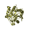
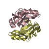
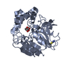
| Title | The structure of the AXH domain of ataxin-1. | ||||||
 Components Components | ATAXIN-1 | ||||||
 Keywords Keywords | RNA BINDING PROTEIN / RNA BINDING / OB-FOLD / HIGH MOBILITY GROUP HOMOLOGY / HMG / DIMERIZATION | ||||||
| Function / homology |  Function and homology information Function and homology informationpoly(G) binding / negative regulation of insulin-like growth factor receptor signaling pathway / POZ domain binding / nuclear inclusion body / nuclear export / poly(U) RNA binding / lung alveolus development / positive regulation of glial cell proliferation / social behavior / RNA processing ...poly(G) binding / negative regulation of insulin-like growth factor receptor signaling pathway / POZ domain binding / nuclear inclusion body / nuclear export / poly(U) RNA binding / lung alveolus development / positive regulation of glial cell proliferation / social behavior / RNA processing / insulin-like growth factor receptor signaling pathway / learning / adult locomotory behavior / excitatory postsynaptic potential / brain development / visual learning / nuclear matrix / memory / transcription by RNA polymerase II / postsynapse / negative regulation of DNA-templated transcription / chromatin binding / nucleolus / negative regulation of transcription by RNA polymerase II / positive regulation of transcription by RNA polymerase II / protein-containing complex / DNA binding / RNA binding / nucleoplasm / identical protein binding / nucleus / cytoplasm / cytosol Similarity search - Function | ||||||
| Biological species |  HOMO SAPIENS (human) HOMO SAPIENS (human) | ||||||
| Method |  X-RAY DIFFRACTION / SYNCHROTRON / MOLECULAR REPLACEMENT / Resolution: 2.5 Å X-RAY DIFFRACTION / SYNCHROTRON / MOLECULAR REPLACEMENT / Resolution: 2.5 Å | ||||||
 Authors Authors | Rees, M. / Chen, Y.W. / de Chiara, C. / Pastore, A. | ||||||
 Citation Citation | Journal: Biophys.J. / Year: 2013 Title: Self-Assembly and Conformational Heterogeneity of the Axh Domain of Ataxin-1: An Unusual Example of a Chameleon Fold Authors: De Chiara, C. / Rees, M. / Menon, R.P. / Pauwels, K. / Lawrence, C. / Konarev, P.V. / Svergun, D.I. / Martin, S.R. / Chen, Y.W. / Pastore, A. #1: Journal: J.Biol.Chem. / Year: 2004Title: The Structure of the Axh Domain of Spinocerebellar Ataxin-1. Authors: Chen, Y.W. / Allen, M.D. / Veprintsev, D.B. / Lowe, J. / Bycroft, M. #2: Journal: FEBS Lett. / Year: 2003 Title: The Axh Module: An Independently Folded Domain Common to Ataxin-1 and Hbp1. Authors: De Chiara, C. / Giannini, C. / Adinolfi, S. / De Boer, J. / Guida, S. / Ramos, A. / Jodice, C. / Kioussis, D. / Pastore, A. #3: Journal: J.Mol.Biol. / Year: 2005 Title: Polyglutamine is not All: The Functional Role of the Axh Domain in the Ataxin-1 Protein. Authors: De Chiara, C. / Menon, R.P. / Dal Piaz, F. / Calder, L. / Pastore, A. | ||||||
| History |
|
- Structure visualization
| Structure viewer | Molecule: MolmilJmol/JSmol |
|---|
- Downloads & links
-Download
| PDBx/mmCIF format | 4apt.cif.gz | 208.1 KB | Display | PDBx/mmCIF format |
|---|---|---|---|---|
| PDB format | pdb4apt.ent.gz | 168.2 KB | Display | PDB format |
| PDBx/mmJSON format | 4apt.json.gz | Tree view | PDBx/mmJSON format | |
| Others |  Other downloads Other downloads |
-Validation report
| Arichive directory | https://data.pdbj.org/pub/pdb/validation_reports/ap/4aptftp://data.pdbj.org/pub/pdb/validation_reports/ap/4apt | HTTPS FTP |
|---|
-Related structure data
| Related structure data | 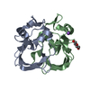 4aqpC 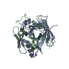 1oa8S S: Starting model for refinement C: citing same article ( |
|---|---|
| Similar structure data |


-Links
 PDBj
PDBj- Assembly
| Deposited unit | 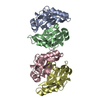
| ||||||||||||
|---|---|---|---|---|---|---|---|---|---|---|---|---|---|
| 1 | 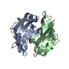
| ||||||||||||
| 2 | 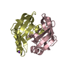
| ||||||||||||
| Unit cell |
| ||||||||||||
| Noncrystallographic symmetry (NCS) | NCS oper:
|
-Components
| #1: Protein | Mass: 13776.670 Da / Num. of mol.: 4 / Fragment: AXH DOMAIN, RESIDUES 566-688 Source method: isolated from a genetically manipulated source Source: (gene. exp.) HOMO SAPIENS (human) / Plasmid: PETM30 / Production host:  #2: Chemical | ChemComp-NA / |   Mass: 22.990 Da / Num. of mol.: 1 / Source method: obtained synthetically / Formula: Na Mass: 22.990 Da / Num. of mol.: 1 / Source method: obtained synthetically / Formula: Na#3: Water | ChemComp-HOH / |  Mass: 18.015 Da / Num. of mol.: 45 / Source method: isolated from a natural source / Formula: H2O Mass: 18.015 Da / Num. of mol.: 45 / Source method: isolated from a natural source / Formula: H2O |
|---|
-Experimental details
-Experiment
| Experiment | Method: X-RAY DIFFRACTION / Number of used crystals: 1 |
|---|
- Sample preparation
| Crystal | Density Matthews: 2.13 Å3/Da / Density % sol: 42.3 % Description: DATA WERE MODIFIED AT THE UCLA DIFFRACTION ANISOTROPY SERVER WITH TRUNCATION ALONG D1, D2, D3 BEING 2. 90,2.50,2.50 ANGSTROMS. ALL VALUES REPORTED ARE AFTER CORRECTION, EXCEPT FOR R- ...Description: DATA WERE MODIFIED AT THE UCLA DIFFRACTION ANISOTROPY SERVER WITH TRUNCATION ALONG D1, D2, D3 BEING 2. 90,2.50,2.50 ANGSTROMS. ALL VALUES REPORTED ARE AFTER CORRECTION, EXCEPT FOR R-MERGE AND DATA REDUNDANCY WHICH ARE FOR UNCORRECTED DATA. |
|---|---|
| Crystal grow | Details: PROTEIN SAMPLES AT 20 MG/ML CRYSTALLISED IN 0.4 M POTASSIUM SODIUM TARTRATE AT ROOM TEMPERATURE. |
-Data collection
| Diffraction | Mean temperature: 100 K |
|---|---|
| Diffraction source | Source: SYNCHROTRON / Site: Diamond  / Beamline: I02 / Wavelength: 0.9796 / Beamline: I02 / Wavelength: 0.9796 |
| Detector | Type: ADSC CCD / Detector: CCD / Date: Dec 7, 2008 |
| Radiation | Protocol: SINGLE WAVELENGTH / Monochromatic (M) / Laue (L): M / Scattering type: x-ray |
| Radiation wavelength | Wavelength: 0.9796 Å / Relative weight: 1 |
| Reflection | Resolution: 2.5→40.4 Å / Num. obs: 14350 / % possible obs: 84.4 % / Observed criterion σ(I): 0 / Redundancy: 4.2 % / Biso Wilson estimate: 42.7 Å2 / Rmerge(I) obs: 0.06 / Net I/σ(I): 12.5 |
| Reflection shell | Resolution: 2.5→2.64 Å / Redundancy: 4.3 % / Rmerge(I) obs: 0.61 / Mean I/σ(I) obs: 3 / % possible all: 35.5 |
- Processing
| Software |
| ||||||||||||||||||||||||||||||||||||||||||||||||||||||||||||||||||||||||||||||||||||||||||||||||||||||||||||||||||||||||||||||||||||||||||||||||||||||||||||||||||||||||||||||||||||||||||||||||||||||||||||||||||||||||||||||||||||||||||||||||||||||||||||||||||||||||||||||||||||||||||||||||||||||||||||
|---|---|---|---|---|---|---|---|---|---|---|---|---|---|---|---|---|---|---|---|---|---|---|---|---|---|---|---|---|---|---|---|---|---|---|---|---|---|---|---|---|---|---|---|---|---|---|---|---|---|---|---|---|---|---|---|---|---|---|---|---|---|---|---|---|---|---|---|---|---|---|---|---|---|---|---|---|---|---|---|---|---|---|---|---|---|---|---|---|---|---|---|---|---|---|---|---|---|---|---|---|---|---|---|---|---|---|---|---|---|---|---|---|---|---|---|---|---|---|---|---|---|---|---|---|---|---|---|---|---|---|---|---|---|---|---|---|---|---|---|---|---|---|---|---|---|---|---|---|---|---|---|---|---|---|---|---|---|---|---|---|---|---|---|---|---|---|---|---|---|---|---|---|---|---|---|---|---|---|---|---|---|---|---|---|---|---|---|---|---|---|---|---|---|---|---|---|---|---|---|---|---|---|---|---|---|---|---|---|---|---|---|---|---|---|---|---|---|---|---|---|---|---|---|---|---|---|---|---|---|---|---|---|---|---|---|---|---|---|---|---|---|---|---|---|---|---|---|---|---|---|---|---|---|---|---|---|---|---|---|---|---|---|---|---|---|---|---|---|---|---|---|---|---|---|---|---|---|---|---|---|---|---|---|---|---|---|---|---|---|---|---|---|---|---|---|---|---|---|---|---|---|
| Refinement | Method to determine structure: MOLECULAR REPLACEMENT Starting model: PDB ENTRY 1OA8 Resolution: 2.5→40.406 Å / SU ML: 0.88 / σ(F): 0 / Phase error: 30.48 / Stereochemistry target values: ML Details: THERE ARE 4 CHEMICALLY IDENTICAL PROTEIN MOLECULES IN THE ASYMMETRIC UNIT. CHAINS A AND B CONSTITUTE A GLOBULAR DIMER, C & D FORM ANOTHER. CHAINS A AND B ARE STRUCTURALLY SLIGHTLY DIFFERENT, ...Details: THERE ARE 4 CHEMICALLY IDENTICAL PROTEIN MOLECULES IN THE ASYMMETRIC UNIT. CHAINS A AND B CONSTITUTE A GLOBULAR DIMER, C & D FORM ANOTHER. CHAINS A AND B ARE STRUCTURALLY SLIGHTLY DIFFERENT, LIKEWISE FOR C & D CHAINS. A AND C ARE MORE ALIKE AND SO ARE CHAINS B & D. DISORDERED REGIONS WERE MODELED STEREOCHEMICALLY
| ||||||||||||||||||||||||||||||||||||||||||||||||||||||||||||||||||||||||||||||||||||||||||||||||||||||||||||||||||||||||||||||||||||||||||||||||||||||||||||||||||||||||||||||||||||||||||||||||||||||||||||||||||||||||||||||||||||||||||||||||||||||||||||||||||||||||||||||||||||||||||||||||||||||||||||
| Solvent computation | Shrinkage radii: 0.6 Å / VDW probe radii: 0.9 Å / Solvent model: FLAT BULK SOLVENT MODEL / Bsol: 34.866 Å2 / ksol: 0.345 e/Å3 | ||||||||||||||||||||||||||||||||||||||||||||||||||||||||||||||||||||||||||||||||||||||||||||||||||||||||||||||||||||||||||||||||||||||||||||||||||||||||||||||||||||||||||||||||||||||||||||||||||||||||||||||||||||||||||||||||||||||||||||||||||||||||||||||||||||||||||||||||||||||||||||||||||||||||||||
| Displacement parameters |
| ||||||||||||||||||||||||||||||||||||||||||||||||||||||||||||||||||||||||||||||||||||||||||||||||||||||||||||||||||||||||||||||||||||||||||||||||||||||||||||||||||||||||||||||||||||||||||||||||||||||||||||||||||||||||||||||||||||||||||||||||||||||||||||||||||||||||||||||||||||||||||||||||||||||||||||
| Refinement step | Cycle: LAST / Resolution: 2.5→40.406 Å
| ||||||||||||||||||||||||||||||||||||||||||||||||||||||||||||||||||||||||||||||||||||||||||||||||||||||||||||||||||||||||||||||||||||||||||||||||||||||||||||||||||||||||||||||||||||||||||||||||||||||||||||||||||||||||||||||||||||||||||||||||||||||||||||||||||||||||||||||||||||||||||||||||||||||||||||
| Refine LS restraints |
| ||||||||||||||||||||||||||||||||||||||||||||||||||||||||||||||||||||||||||||||||||||||||||||||||||||||||||||||||||||||||||||||||||||||||||||||||||||||||||||||||||||||||||||||||||||||||||||||||||||||||||||||||||||||||||||||||||||||||||||||||||||||||||||||||||||||||||||||||||||||||||||||||||||||||||||
| LS refinement shell |
| ||||||||||||||||||||||||||||||||||||||||||||||||||||||||||||||||||||||||||||||||||||||||||||||||||||||||||||||||||||||||||||||||||||||||||||||||||||||||||||||||||||||||||||||||||||||||||||||||||||||||||||||||||||||||||||||||||||||||||||||||||||||||||||||||||||||||||||||||||||||||||||||||||||||||||||
| Refinement TLS params. | Method: refined / Refine-ID: X-RAY DIFFRACTION
| ||||||||||||||||||||||||||||||||||||||||||||||||||||||||||||||||||||||||||||||||||||||||||||||||||||||||||||||||||||||||||||||||||||||||||||||||||||||||||||||||||||||||||||||||||||||||||||||||||||||||||||||||||||||||||||||||||||||||||||||||||||||||||||||||||||||||||||||||||||||||||||||||||||||||||||
| Refinement TLS group |
|
