Movie
Movie Controller

[English] 日本語
 Yorodumi
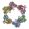
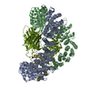
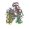
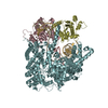
- PDB-6w4x: Holocomplex of E. coli class Ia ribonucleotide reductase with GDP... -
+ Open data

- Basic information
| Entry | Database: PDB / ID: 6w4x | ||||||
|---|---|---|---|---|---|---|---|
| Title | Holocomplex of E. coli class Ia ribonucleotide reductase with GDP and TTP | ||||||
 Components Components | (Ribonucleoside-diphosphate reductase 1 subunit ...) x 2 | ||||||
 Keywords Keywords | OXIDOREDUCTASE / complex | ||||||
| Function / homology |  Function and homology information Function and homology informationribonucleoside diphosphate metabolic process / 2'-deoxyribonucleotide biosynthetic process / nucleobase-containing small molecule interconversion / ribonucleoside-diphosphate reductase complex / ribonucleoside-diphosphate reductase / ribonucleoside-diphosphate reductase activity, thioredoxin disulfide as acceptor / deoxyribonucleotide biosynthetic process / protein folding chaperone / iron ion binding / ATP binding ...ribonucleoside diphosphate metabolic process / 2'-deoxyribonucleotide biosynthetic process / nucleobase-containing small molecule interconversion / ribonucleoside-diphosphate reductase complex / ribonucleoside-diphosphate reductase / ribonucleoside-diphosphate reductase activity, thioredoxin disulfide as acceptor / deoxyribonucleotide biosynthetic process / protein folding chaperone / iron ion binding / ATP binding / identical protein binding / cytoplasm / cytosol Similarity search - Function | ||||||
| Biological species |  | ||||||
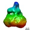
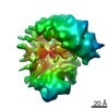
| Method | ELECTRON MICROSCOPY / single particle reconstruction / cryo EM / Resolution: 3.6 Å | ||||||
 Authors Authors | Kang, G. / Taguchi, A. / Stubbe, J. / Drennan, C.L. | ||||||
| Funding support |  United States, 1items United States, 1items
| ||||||
 Citation Citation | Journal: Science / Year: 2020 Title: Structure of a trapped radical transfer pathway within a ribonucleotide reductase holocomplex. Authors: Gyunghoon Kang / Alexander T Taguchi / JoAnne Stubbe / Catherine L Drennan / Abstract: Ribonucleotide reductases (RNRs) are a diverse family of enzymes that are alone capable of generating 2'-deoxynucleotides de novo and are thus critical in DNA biosynthesis and repair. The nucleotide ...Ribonucleotide reductases (RNRs) are a diverse family of enzymes that are alone capable of generating 2'-deoxynucleotides de novo and are thus critical in DNA biosynthesis and repair. The nucleotide reduction reaction in all RNRs requires the generation of a transient active site thiyl radical, and in class I RNRs, this process involves a long-range radical transfer between two subunits, α and β. Because of the transient subunit association, an atomic resolution structure of an active α2β2 RNR complex has been elusive. We used a doubly substituted β2, E52Q/(2,3,5)-trifluorotyrosine122-β2, to trap wild-type α2 in a long-lived α2β2 complex. We report the structure of this complex by means of cryo-electron microscopy to 3.6-angstrom resolution, allowing for structural visualization of a 32-angstrom-long radical transfer pathway that affords RNR activity. | ||||||
| History |
|
- Structure visualization
| Movie |
Movie viewer |
|---|---|
| Structure viewer | Molecule: MolmilJmol/JSmol |
- Downloads & links
-Download
| PDBx/mmCIF format | 6w4x.cif.gz | 394 KB | Display | PDBx/mmCIF format |
|---|---|---|---|---|
| PDB format | pdb6w4x.ent.gz | 319.7 KB | Display | PDB format |
| PDBx/mmJSON format | 6w4x.json.gz | Tree view | PDBx/mmJSON format | |
| Others |  Other downloads Other downloads |
-Validation report
| Arichive directory | https://data.pdbj.org/pub/pdb/validation_reports/w4/6w4xftp://data.pdbj.org/pub/pdb/validation_reports/w4/6w4x | HTTPS FTP |
|---|
-Related structure data
| Related structure data |  21540MC M: map data used to model this data C: citing same article ( |
|---|---|
| Similar structure data |
-Links
 PDBj
PDBj
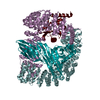
- Assembly
| Deposited unit | 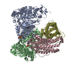
|
|---|---|
| 1 |
|
-Components
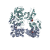
-Ribonucleoside-diphosphate reductase 1 subunit ... , 2 types, 4 molecules ABCD
| #1: Protein | Mass: 85877.086 Da / Num. of mol.: 2 Source method: isolated from a genetically manipulated source Source: (gene. exp.) Strain: K12 / Gene: nrdA, dnaF, b2234, JW2228 / Production host: References: UniProt: P00452, ribonucleoside-diphosphate reductase #2: Protein | Mass: 43611.039 Da / Num. of mol.: 2 Source method: isolated from a genetically manipulated source Source: (gene. exp.) Strain: K12 / Gene: nrdB, ftsB, b2235, JW2229 / Production host: References: UniProt: P69924, ribonucleoside-diphosphate reductase |
|---|
-Non-polymers , 5 types, 11 molecules 


| #3: Chemical |  Mass: 482.168 Da / Num. of mol.: 2 / Source method: obtained synthetically / Formula: C10H17N2O14P3 Mass: 482.168 Da / Num. of mol.: 2 / Source method: obtained synthetically / Formula: C10H17N2O14P3#4: Chemical |  Mass: 24.305 Da / Num. of mol.: 2 / Source method: obtained synthetically / Formula: Mg Mass: 24.305 Da / Num. of mol.: 2 / Source method: obtained synthetically / Formula: Mg#5: Chemical | ChemComp-GDP / |  Type: RNA linking / Mass: 443.201 Da / Num. of mol.: 1 / Source method: obtained synthetically / Formula: C10H15N5O11P2 / Comment: GDP, energy-carrying molecule*YM Type: RNA linking / Mass: 443.201 Da / Num. of mol.: 1 / Source method: obtained synthetically / Formula: C10H15N5O11P2 / Comment: GDP, energy-carrying molecule*YM#6: Chemical |  Mass: 127.689 Da / Num. of mol.: 2 / Source method: obtained synthetically / Formula: Fe2O Mass: 127.689 Da / Num. of mol.: 2 / Source method: obtained synthetically / Formula: Fe2O#7: Water | ChemComp-HOH / | Mass: 18.015 Da / Num. of mol.: 4 / Source method: isolated from a natural source / Formula: H2O |
|---|
-Details
| Has ligand of interest | Y |
|---|
-Experimental details
-Experiment
| Experiment | Method: ELECTRON MICROSCOPY |
|---|---|
| EM experiment | Aggregation state: PARTICLE / 3D reconstruction method: single particle reconstruction |
- Sample preparation
| Component | Name: Holocomplex of E. coli class Ia ribonucleotide reductase with GDP and TTP Type: COMPLEX / Entity ID: #1-#2 / Source: RECOMBINANT |
|---|---|
| Source (natural) | Organism: |
| Source (recombinant) | Organism: |
| Buffer solution | pH: 7.6 |
| Specimen | Embedding applied: NO / Shadowing applied: NO / Staining applied: NO / Vitrification applied: YES |
| Specimen support | Grid material: COPPER / Grid mesh size: 300 divisions/in. / Grid type: Quantifoil R1.2/1.3 |
| Vitrification | Instrument: FEI VITROBOT MARK IV / Cryogen name: ETHANE / Humidity: 90 % / Chamber temperature: 283 K / Details: blot for 4.5 seconds before plunging |
- Electron microscopy imaging
| Experimental equipment |  Model: Titan Krios / Image courtesy: FEI Company |
|---|---|
| Microscopy | Model: FEI TITAN KRIOS |
| Electron gun | Electron source:  FIELD EMISSION GUN / Accelerating voltage: 300 kV / Illumination mode: FLOOD BEAM FIELD EMISSION GUN / Accelerating voltage: 300 kV / Illumination mode: FLOOD BEAM |
| Electron lens | Mode: BRIGHT FIELD |
| Image recording | Average exposure time: 6 sec. / Electron dose: 47.1 e/Å2 / Film or detector model: GATAN K2 SUMMIT (4k x 4k) / Num. of grids imaged: 2 / Num. of real images: 6238 |
| Image scans | Movie frames/image: 30 |
- Processing
| Software | Name: PHENIX / Version: 1.15.2_3472: / Classification: refinement | ||||||||||||||||||||||||||||||||||||
|---|---|---|---|---|---|---|---|---|---|---|---|---|---|---|---|---|---|---|---|---|---|---|---|---|---|---|---|---|---|---|---|---|---|---|---|---|---|
| EM software |
| ||||||||||||||||||||||||||||||||||||
| CTF correction | Type: PHASE FLIPPING AND AMPLITUDE CORRECTION | ||||||||||||||||||||||||||||||||||||
| Symmetry | Point symmetry: C1 (asymmetric) | ||||||||||||||||||||||||||||||||||||
| 3D reconstruction | Resolution: 3.6 Å / Resolution method: FSC 0.143 CUT-OFF / Num. of particles: 80386 / Symmetry type: POINT | ||||||||||||||||||||||||||||||||||||
| Atomic model building | Space: REAL | ||||||||||||||||||||||||||||||||||||
| Atomic model building | PDB-ID: 5CNV Accession code: 5CNV / Source name: PDB / Type: experimental model | ||||||||||||||||||||||||||||||||||||
| Refine LS restraints |
|