 Movie
Movie Controller

[English] 日本語
 Yorodumi
- PDB-6og1: Focus classification structure of the hyperactive ClpB mutant K47... -
+ Open data

- Basic information
| Entry | Database: PDB / ID: 6og1 | ||||||
|---|---|---|---|---|---|---|---|
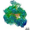
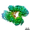
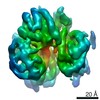
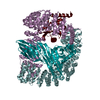
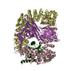
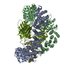
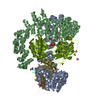
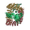
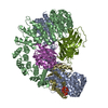
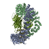
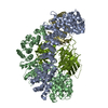
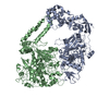
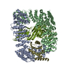
| Title | Focus classification structure of the hyperactive ClpB mutant K476C, bound to casein, pre-state | ||||||
 Components Components | Hyperactive disaggregase ClpB | ||||||
 Keywords Keywords | CHAPERONE / disaggregase / CLPB / AAA+ | ||||||
| Function / homology |  Function and homology information Function and homology informationcellular response to heat / response to heat / protein refolding / ATP hydrolysis activity / ATP binding / membrane / identical protein binding / cytoplasm / cytosol Similarity search - Function | ||||||
| Biological species |  | ||||||
| Method | ELECTRON MICROSCOPY / single particle reconstruction / cryo EM / Resolution: 3.3 Å | ||||||
 Authors Authors | Rizo, A.R. / Lin, J.-B. / Gates, S.N. / Tse, E. / Bart, S.M. / Castellano, L.M. / Dimaio, F. / Shorter, J. / Southworth, D.R. | ||||||
 Citation Citation | Journal: Nat Commun / Year: 2019 Title: Structural basis for substrate gripping and translocation by the ClpB AAA+ disaggregase. Authors: Alexandrea N Rizo / JiaBei Lin / Stephanie N Gates / Eric Tse / Stephen M Bart / Laura M Castellano / Frank DiMaio / James Shorter / Daniel R Southworth /  Abstract: Bacterial ClpB and yeast Hsp104 are homologous Hsp100 protein disaggregases that serve critical functions in proteostasis by solubilizing protein aggregates. Two AAA+ nucleotide binding domains (NBDs) ...Bacterial ClpB and yeast Hsp104 are homologous Hsp100 protein disaggregases that serve critical functions in proteostasis by solubilizing protein aggregates. Two AAA+ nucleotide binding domains (NBDs) power polypeptide translocation through a central channel comprised of a hexameric spiral of protomers that contact substrate via conserved pore-loop interactions. Here we report cryo-EM structures of a hyperactive ClpB variant bound to the model substrate, casein in the presence of slowly hydrolysable ATPγS, which reveal the translocation mechanism. Distinct substrate-gripping interactions are identified for NBD1 and NBD2 pore loops. A trimer of N-terminal domains define a channel entrance that binds the polypeptide substrate adjacent to the topmost NBD1 contact. NBD conformations at the seam interface reveal how ATP hydrolysis-driven substrate disengagement and re-binding are precisely tuned to drive a directional, stepwise translocation cycle. | ||||||
| History |
| ||||||
| Remark 650 | HELIX DETERMINATION METHOD: AUTHOR | ||||||
| Remark 700 | SHEET DETERMINATION METHOD: AUTHOR |
- Structure visualization
| Movie |
Movie viewer |
|---|---|
| Structure viewer | Molecule: MolmilJmol/JSmol |
- Downloads & links
-Download
| PDBx/mmCIF format | 6og1.cif.gz | 217.5 KB | Display | PDBx/mmCIF format |
|---|---|---|---|---|
| PDB format | pdb6og1.ent.gz | 169.4 KB | Display | PDB format |
| PDBx/mmJSON format | 6og1.json.gz | Tree view | PDBx/mmJSON format | |
| Others |  Other downloads Other downloads |
-Validation report
| Arichive directory | https://data.pdbj.org/pub/pdb/validation_reports/og/6og1ftp://data.pdbj.org/pub/pdb/validation_reports/og/6og1 | HTTPS FTP |
|---|
-Related structure data
| Related structure data |  20049MC  6oaxC  6oayC  6og2C 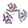 6og3C M: map data used to model this data C: citing same article ( |
|---|---|
| Similar structure data |
-Links
 PDBj
PDBj


- Assembly
| Deposited unit | 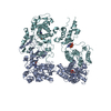
|
|---|---|
| 1 |
|
-Components
| #1: Protein | Mass: 97018.469 Da / Num. of mol.: 2 / Mutation: K476C Source method: isolated from a genetically manipulated source Source: (gene. exp.) #2: Chemical |   Mass: 427.201 Da / Num. of mol.: 3 / Source method: obtained synthetically / Formula: C10H15N5O10P2 / Comment: ADP, energy-carrying molecule*YM Mass: 427.201 Da / Num. of mol.: 3 / Source method: obtained synthetically / Formula: C10H15N5O10P2 / Comment: ADP, energy-carrying molecule*YM#3: Chemical | ChemComp-AGS / |   Mass: 523.247 Da / Num. of mol.: 1 / Source method: obtained synthetically / Formula: C10H16N5O12P3S / Comment: ATP-gamma-S, energy-carrying molecule analogue*YM Mass: 523.247 Da / Num. of mol.: 1 / Source method: obtained synthetically / Formula: C10H16N5O12P3S / Comment: ATP-gamma-S, energy-carrying molecule analogue*YM |
|---|
-Experimental details
-Experiment
| Experiment | Method: ELECTRON MICROSCOPY |
|---|---|
| EM experiment | Aggregation state: PARTICLE / 3D reconstruction method: single particle reconstruction |
- Sample preparation
| Component | Name: hyperactive disaggregase ClpB K476C, bound to casein / Type: COMPLEX / Entity ID: #1 / Source: RECOMBINANT |
|---|---|
| Molecular weight | Experimental value: YES |
| Source (natural) | Organism: |
| Source (recombinant) | Organism: |
| Buffer solution | pH: 7.4 |
| Specimen | Embedding applied: NO / Shadowing applied: NO / Staining applied: NO / Vitrification applied: YES |
| Specimen support | Details: unspecified |
| Vitrification | Cryogen name: ETHANE |
- Electron microscopy imaging
| Experimental equipment |  Model: Titan Krios / Image courtesy: FEI Company |
|---|---|
| Microscopy | Model: FEI TITAN KRIOS |
| Electron gun | Electron source:  FIELD EMISSION GUN / Accelerating voltage: 300 kV / Illumination mode: FLOOD BEAM FIELD EMISSION GUN / Accelerating voltage: 300 kV / Illumination mode: FLOOD BEAM |
| Electron lens | Mode: BRIGHT FIELD / C2 aperture diameter: 2.6 µm |
| Image recording | Electron dose: 56 e/Å2 / Film or detector model: GATAN K2 SUMMIT (4k x 4k) |
- Processing
| CTF correction | Type: PHASE FLIPPING AND AMPLITUDE CORRECTION |
|---|---|
| Symmetry | Point symmetry: C1 (asymmetric) |
| 3D reconstruction | Resolution: 3.3 Å / Resolution method: FSC 0.143 CUT-OFF / Num. of particles: 221083 / Symmetry type: POINT |
