Movie
Movie Controller

[English] 日本語
 Yorodumi
- EMDB-21540: Holocomplex of E. coli class Ia ribonucleotide reductase with GDP... -
+ Open data

- Basic information
| Entry | Database: EMDB / ID: EMD-21540 | |||||||||
|---|---|---|---|---|---|---|---|---|---|---|
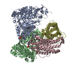
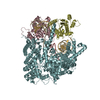
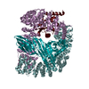
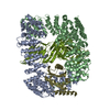
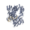
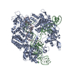
| Title | Holocomplex of E. coli class Ia ribonucleotide reductase with GDP and TTP | |||||||||
 Map data Map data | Holocomplex of E. coli class Ia ribonucleotide reductase with GDP and TTP | |||||||||
 Sample Sample |
| |||||||||
 Keywords Keywords | complex / OXIDOREDUCTASE | |||||||||
| Function / homology |  Function and homology information Function and homology informationribonucleoside diphosphate metabolic process / 2'-deoxyribonucleotide biosynthetic process / nucleobase-containing small molecule interconversion / ribonucleoside-diphosphate reductase complex / ribonucleoside-diphosphate reductase / ribonucleoside-diphosphate reductase activity, thioredoxin disulfide as acceptor / deoxyribonucleotide biosynthetic process / protein folding chaperone / iron ion binding / ATP binding ...ribonucleoside diphosphate metabolic process / 2'-deoxyribonucleotide biosynthetic process / nucleobase-containing small molecule interconversion / ribonucleoside-diphosphate reductase complex / ribonucleoside-diphosphate reductase / ribonucleoside-diphosphate reductase activity, thioredoxin disulfide as acceptor / deoxyribonucleotide biosynthetic process / protein folding chaperone / iron ion binding / ATP binding / identical protein binding / cytoplasm / cytosol Similarity search - Function | |||||||||
| Biological species |  | |||||||||
| Method | single particle reconstruction / cryo EM / Resolution: 3.6 Å | |||||||||
 Authors Authors | Kang G / Taguchi A | |||||||||
| Funding support |  United States, 1 items United States, 1 items
| |||||||||
 Citation Citation | Journal: Science / Year: 2020 Title: Structure of a trapped radical transfer pathway within a ribonucleotide reductase holocomplex. Authors: Gyunghoon Kang / Alexander T Taguchi / JoAnne Stubbe / Catherine L Drennan / Abstract: Ribonucleotide reductases (RNRs) are a diverse family of enzymes that are alone capable of generating 2'-deoxynucleotides de novo and are thus critical in DNA biosynthesis and repair. The nucleotide ...Ribonucleotide reductases (RNRs) are a diverse family of enzymes that are alone capable of generating 2'-deoxynucleotides de novo and are thus critical in DNA biosynthesis and repair. The nucleotide reduction reaction in all RNRs requires the generation of a transient active site thiyl radical, and in class I RNRs, this process involves a long-range radical transfer between two subunits, α and β. Because of the transient subunit association, an atomic resolution structure of an active α2β2 RNR complex has been elusive. We used a doubly substituted β2, E52Q/(2,3,5)-trifluorotyrosine122-β2, to trap wild-type α2 in a long-lived α2β2 complex. We report the structure of this complex by means of cryo-electron microscopy to 3.6-angstrom resolution, allowing for structural visualization of a 32-angstrom-long radical transfer pathway that affords RNR activity. | |||||||||
| History |
|
- Structure visualization
| Movie |
Movie viewer |
|---|---|
| Structure viewer | EM map: SurfViewMolmilJmol/JSmol |
| Supplemental images |

- Downloads & links
-EMDB archive
| Map data | emd_21540.map.gz | 49.3 MB | EMDB map data format | |
|---|---|---|---|---|
| Header (meta data) | emd-21540-v30.xmlemd-21540.xml | 19.2 KB 19.2 KB | Display Display | EMDB header |
| FSC (resolution estimation) | emd_21540_fsc.xml | 8.6 KB | Display | FSC data file |
| Images |  emd_21540.png emd_21540.png | 77.8 KB | ||
| Filedesc metadata | emd-21540.cif.gz | 6.6 KB | ||
| Others | emd_21540_half_map_1.map.gzemd_21540_half_map_2.map.gz | 40.8 MB 40.8 MB | ||
| Archive directory |  http://ftp.pdbj.org/pub/emdb/structures/EMD-21540ftp://ftp.pdbj.org/pub/emdb/structures/EMD-21540 http://ftp.pdbj.org/pub/emdb/structures/EMD-21540ftp://ftp.pdbj.org/pub/emdb/structures/EMD-21540 | HTTPS FTP |
-Related structure data
| Related structure data | 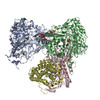 6w4xMC M: atomic model generated by this map C: citing same article ( |
|---|---|
| Similar structure data |


-Links
| EMDB pages | EMDB (EBI/PDBe) / EMDataResource |
|---|---|
| Related items in Molecule of the Month |

-Map
| File | Download / File: emd_21540.map.gz / Format: CCP4 / Size: 52.7 MB / Type: IMAGE STORED AS FLOATING POINT NUMBER (4 BYTES) | ||||||||||||||||||||||||||||||||||||||||||||||||||||||||||||||||||||
|---|---|---|---|---|---|---|---|---|---|---|---|---|---|---|---|---|---|---|---|---|---|---|---|---|---|---|---|---|---|---|---|---|---|---|---|---|---|---|---|---|---|---|---|---|---|---|---|---|---|---|---|---|---|---|---|---|---|---|---|---|---|---|---|---|---|---|---|---|---|
| Annotation | Holocomplex of E. coli class Ia ribonucleotide reductase with GDP and TTP | ||||||||||||||||||||||||||||||||||||||||||||||||||||||||||||||||||||
| Projections & slices | Image control
Images are generated by Spider. | ||||||||||||||||||||||||||||||||||||||||||||||||||||||||||||||||||||
| Voxel size | X=Y=Z: 1.059 Å | ||||||||||||||||||||||||||||||||||||||||||||||||||||||||||||||||||||
| Density |
| ||||||||||||||||||||||||||||||||||||||||||||||||||||||||||||||||||||
| Symmetry | Space group: 1 | ||||||||||||||||||||||||||||||||||||||||||||||||||||||||||||||||||||
| Details | EMDB XML:
CCP4 map header:
| ||||||||||||||||||||||||||||||||||||||||||||||||||||||||||||||||||||
 Z (Sec.)
 Y (Row.)
 X (Col.)


-Supplemental data
-Half map: #1
| File | emd_21540_half_map_1.map | ||||||||||||
|---|---|---|---|---|---|---|---|---|---|---|---|---|---|
| Projections & Slices |
| ||||||||||||
| Density Histograms |


-Half map: #2
| File | emd_21540_half_map_2.map | ||||||||||||
|---|---|---|---|---|---|---|---|---|---|---|---|---|---|
| Projections & Slices |
| ||||||||||||
| Density Histograms |


- Sample components
-Entire : Holocomplex of E. coli class Ia ribonucleotide reductase with GDP...
| Entire | Name: Holocomplex of E. coli class Ia ribonucleotide reductase with GDP and TTP |
|---|---|
| Components |
|
-Supramolecule #1: Holocomplex of E. coli class Ia ribonucleotide reductase with GDP...
| Supramolecule | Name: Holocomplex of E. coli class Ia ribonucleotide reductase with GDP and TTP type: complex / ID: 1 / Parent: 0 / Macromolecule list: #1-#2 |
|---|---|
| Source (natural) | Organism: |
-Macromolecule #1: Ribonucleoside-diphosphate reductase 1 subunit alpha
| Macromolecule | Name: Ribonucleoside-diphosphate reductase 1 subunit alpha / type: protein_or_peptide / ID: 1 / Number of copies: 2 / Enantiomer: LEVO / EC number: ribonucleoside-diphosphate reductase |
|---|---|
| Source (natural) | Organism: |
| Molecular weight | Theoretical: 85.877086 KDa |
| Recombinant expression | Organism: |
| Sequence | String: MNQNLLVTKR DGSTERINLD KIHRVLDWAA EGLHNVSISQ VELRSHIQFY DGIKTSDIHE TIIKAAADLI SRDAPDYQYL AARLAIFHL RKKAYGQFEP PALYDHVVKM VEMGKYDNHL LEDYTEEEFK QMDTFIDHDR DMTFSYAAVK QLEGKYLVQN R VTGEIYES ...String: MNQNLLVTKR DGSTERINLD KIHRVLDWAA EGLHNVSISQ VELRSHIQFY DGIKTSDIHE TIIKAAADLI SRDAPDYQYL AARLAIFHL RKKAYGQFEP PALYDHVVKM VEMGKYDNHL LEDYTEEEFK QMDTFIDHDR DMTFSYAAVK QLEGKYLVQN R VTGEIYES AQFLYILVAA CLFSNYPRET RLQYVKRFYD AVSTFKISLP TPIMSGVRTP TRQFSSCVLI ECGDSLDSIN AT SSAIVKY VSQRAGIGIN AGRIRALGSP IRGGEAFHTG CIPFYKHFQT AVKSCSQGGV RGGAATLFYP MWHLEVESLL VLK NNRGVE GNRVRHMDYG VQINKLMYTR LLKGEDITLF SPSDVPGLYD AFFADQEEFE RLYTKYEKDD SIRKQRVKAV ELFS LMMQE RASTGRIYIQ NVDHCNTHSP FDPAIAPVRQ SNLCLEIALP TKPLNDVNDE NGEIALCTLS AFNLGAINNL DELEE LAIL AVRALDALLD YQDYPIPAAK RGAMGRRTLG IGVINFAYYL AKHGKRYSDG SANNLTHKTF EAIQYYLLKA SNELAK EQG ACPWFNETTY AKGILPIDTY KKDLDTIANE PLHYDWEALR ESIKTHGLRN STLSALMPSE TSSQISNATN GIEPPRG YV SIKASKDGIL RQVVPDYEHL HDAYELLWEM PGNDGYLQLV GIMQKFIDQS ISANTNYDPS RFPSGKVPMQ QLLKDLLT A YKFGVKTLYY QNTRDGAEDA QDDLVPSIQD DGCESGACKI UniProtKB: Ribonucleoside-diphosphate reductase 1 subunit alpha |
-Macromolecule #2: Ribonucleoside-diphosphate reductase 1 subunit beta
| Macromolecule | Name: Ribonucleoside-diphosphate reductase 1 subunit beta / type: protein_or_peptide / ID: 2 / Number of copies: 2 / Enantiomer: LEVO / EC number: ribonucleoside-diphosphate reductase |
|---|---|
| Source (natural) | Organism: |
| Molecular weight | Theoretical: 43.611039 KDa |
| Recombinant expression | Organism: |
| Sequence | String: MAYTTFSQTK NDQLKEPMFF GQPVNVARYD QQKYDIFEKL IEKQLSFFWR PEQVDVSRDR IDYQALPEHE KHIFISNLKY QTLLDSIQG RSPNVALLPL ISIPELETWV ETWAFSETIH SRS(FY3)THIIRN IVNDPSVVFD DIVTNEQIQK RAEGISS YY DELIEMTSYW ...String: MAYTTFSQTK NDQLKEPMFF GQPVNVARYD QQKYDIFEKL IEKQLSFFWR PEQVDVSRDR IDYQALPEHE KHIFISNLKY QTLLDSIQG RSPNVALLPL ISIPELETWV ETWAFSETIH SRS(FY3)THIIRN IVNDPSVVFD DIVTNEQIQK RAEGISS YY DELIEMTSYW HLLGEGTHTV NGKTVTVSLR ELKKKLYLCL MSVNALEAIR FYVSFACSFA FAERELMEGN AKIIRLIA R DEALHLTGTQ HMLNLLRSGA DDPEMAEIAE ECKQECYDLF VQAAQQEKDW ADYLFRDGSM IGLNKDILCQ YVEYITNIR MQAVGLDLPF QTRSNPIPWI NTWLVSDNVQ VAPQEVEVSS YLVGQIDSEV DTDDLSNFQL UniProtKB: Ribonucleoside-diphosphate reductase 1 subunit beta |
-Macromolecule #3: THYMIDINE-5'-TRIPHOSPHATE
| Macromolecule | Name: THYMIDINE-5'-TRIPHOSPHATE / type: ligand / ID: 3 / Number of copies: 2 / Formula: TTP |
|---|---|
| Molecular weight | Theoretical: 482.168 Da |
| Chemical component information |  ChemComp-TTP: |
-Macromolecule #4: MAGNESIUM ION
| Macromolecule | Name: MAGNESIUM ION / type: ligand / ID: 4 / Number of copies: 2 / Formula: MG |
|---|---|
| Molecular weight | Theoretical: 24.305 Da |
-Macromolecule #5: GUANOSINE-5'-DIPHOSPHATE
| Macromolecule | Name: GUANOSINE-5'-DIPHOSPHATE / type: ligand / ID: 5 / Number of copies: 1 / Formula: GDP |
|---|---|
| Molecular weight | Theoretical: 443.201 Da |
| Chemical component information |  ChemComp-GDP: |
-Macromolecule #6: MU-OXO-DIIRON
| Macromolecule | Name: MU-OXO-DIIRON / type: ligand / ID: 6 / Number of copies: 2 / Formula: FEO |
|---|---|
| Molecular weight | Theoretical: 127.689 Da |
| Chemical component information |  ChemComp-FEO: |
-Macromolecule #7: water
| Macromolecule | Name: water / type: ligand / ID: 7 / Number of copies: 4 / Formula: HOH |
|---|---|
| Molecular weight | Theoretical: 18.015 Da |
| Chemical component information |  ChemComp-HOH: |
-Experimental details
-Structure determination
| Method | cryo EM |
|---|---|
 Processing Processing | single particle reconstruction |
| Aggregation state | particle |
-Sample preparation
| Buffer | pH: 7.6 |
|---|---|
| Grid | Model: Quantifoil R1.2/1.3 / Material: COPPER / Mesh: 300 / Pretreatment - Type: GLOW DISCHARGE / Pretreatment - Time: 60 sec. |
| Vitrification | Cryogen name: ETHANE / Chamber humidity: 90 % / Chamber temperature: 283 K / Instrument: FEI VITROBOT MARK IV / Details: blot for 4.5 seconds before plunging. |
- Electron microscopy
| Microscope | FEI TITAN KRIOS |
|---|---|
| Image recording | Film or detector model: GATAN K2 SUMMIT (4k x 4k) / Number grids imaged: 2 / Number real images: 6238 / Average exposure time: 6.0 sec. / Average electron dose: 47.1 e/Å2 |
| Electron beam | Acceleration voltage: 300 kV / Electron source:  FIELD EMISSION GUN FIELD EMISSION GUN |
| Electron optics | Illumination mode: FLOOD BEAM / Imaging mode: BRIGHT FIELD |
| Experimental equipment |  Model: Titan Krios / Image courtesy: FEI Company |